Adapting to Technological Shifts in Orthodontic Care

Adapting to Technological Shifts in Orthodontic Care

**Early Intervention with Invisalign First for Kids**

The field of orthodontics has witnessed a remarkable evolution, especially in recent years, as technological advancements have significantly improved treatment options for children. One of the most notable developments is the use of clear aligners, which have become a popular choice for kids due to their comfort, aesthetic appeal, and ease of maintenance. Retainers are often needed after braces to maintain alignment Child-friendly orthodontic solutions malocclusion. Unlike traditional metal braces, clear aligners are removable, allowing for better oral hygiene and reduced discomfort during meals and brushing.


Technological innovations such as artificial intelligence (AI) have further enhanced the effectiveness of clear aligners. AI-powered systems can analyze a child's dental structure and predict the most efficient paths for teeth alignment, leading to customized treatment plans that are more precise and efficient. This approach not only streamlines the treatment process but also reduces the number of visits required to the orthodontist, making it more appealing for parents and children.


Advances in materials science are also transforming the comfort and durability of clear aligners. Future aligners are expected to be made from innovative materials that increase flexibility and durability, ensuring a less intrusive experience for young patients. Additionally, the integration of digital imaging and 3D printing technologies allows for precise customization of orthodontic appliances, ensuring a better fit and more effective treatment outcomes.


The integration of robotics and automation in orthodontics has improved the precision and safety of complex procedures. Virtual reality is also becoming a tool for treatment planning, enabling patients and orthodontists to visualize possible outcomes before starting treatment. This not only helps in making more accurate treatment plans but also enhances patient understanding and confidence in the treatment process.


Innovations like smart braces and self-ligating systems are further optimizing orthodontic care. These systems use specialized technology to monitor tooth movement and provide real-time data, allowing for adjustments that can shorten treatment times and improve results.


The future of orthodontics for kids is poised to become even more personalized and efficient, with technology at the forefront. As these advancements become more available and effective, they will redefine how early orthodontic treatments are approached, making them more appealing and effective for young patients.

The field of orthodontics is experiencing a significant transformation, driven by technological advancements that are revolutionizing treatment options for children. These innovations not only enhance the precision and efficiency of orthodontic care but also offer more comfortable and personalized solutions. At the forefront of these developments is the integration of artificial intelligence (AI) and the use of advanced materials, which are significantly improving the effectiveness of both clear aligners and traditional braces.


AI integration in orthodontics allows for the analysis of vast amounts of patient data, enabling orthodontists to predict treatment outcomes with greater accuracy. This technology helps in crafting personalized treatment plans tailored to each patient's unique dental structure, leading to more efficient treatments and better results. Additionally, AI assists in monitoring treatment progress, providing real-time feedback that allows for timely adjustments and minimizes potential complications.


Advanced materials are also transforming the field by enhancing the comfort and aesthetics of orthodontic appliances. Clear aligners, for instance, have become a popular alternative to traditional metal braces, offering a virtually invisible and removable option that allows for easier maintenance of oral hygiene. The use of 3D printing technology further customizes these appliances, ensuring a precise fit that enhances both comfort and effectiveness.


Furthermore, technological advancements have made orthodontic care more patient-centric. Virtual consultations and remote monitoring technologies allow patients to receive expert advice and track their treatment progress without the need for frequent office visits. This not only saves time but also increases accessibility, making high-quality orthodontic care more convenient for children and their families.


In summary, technological innovations in orthodontics are creating a new era of patient care that is more precise, efficient, and comfortable. As these advancements continue to shape the field, children can look forward to more effective, aesthetically pleasing, and personalized treatment options that enhance their overall dental health and well-being.

**The HealthyStart System**

The advent of artificial intelligence (AI) in orthodontic treatment has revolutionized the field, transforming the way orthodontists diagnose, plan, and execute treatments. This technological shift is not only enhancing the precision and efficiency of orthodontic care but also improving patient outcomes.


At the forefront of these advancements is AI's role in diagnosis. By leveraging machine learning algorithms, AI systems can analyze radiographic images with high accuracy, correcting noise and providing detailed cephalometric reports in minutes. This not only aids in the identification of malocclusions but also facilitates better visualization of treatment outcomes, thereby enhancing decision support for orthodontists[1][3]. AI's ability to quickly and reliable diagnose conditions allows for more personalized treatment plans, which are crucial in orthodontics where every case is unique[1][3]. Moreover, AI-powered software can assist in simulating tooth movements and predicting treatment outcomes, which helps in optimizing treatment sequences[3][4]. This approach is especially significant in managing complex cases where AI can provide guidance to inexperienced orthodontists and support surgical planning for maxillofacial deformities[1][3]. AI also aids in the surgical planning of orthognathic surgeries by providing precise data, which is essential for comprehensive treatment[1]. Furthermore, AI's face-driven treatment planning approach focuses on enhancing facial aesthetics, which is a key aspect of orthodontic care[1][3]. This personalized approach not only results in better clinical outcomes but also enhances patient satisfaction by aligning treatment with individual facial features[1][3]. AI's impact on orthodontics also includes the automated fabrication of orthodontic appliances such as clear aligners and indirect bonding trays. These advancements offer better esthetics, comfort, and acceptability for patients, replacing traditional methods with more precise and customized solutions[1][5]. The use of 3D printing technology, often in alignment with AI, allows for the fabrication of customized brackets and aligners that fit each patient's unique dental and facial features. This not only enhances the treatment's efficiency but also provides a more personalized and effective treatment plan[5]. Another significant aspect of AI in orthodontics is its role in operational efficiency. AI can automatically schedule staff, organize patient appointments, and perform tasks like dental coding and billing, thereby increasing operational efficiency and patient satisfaction[1]. This efficiency also allows for cost-savings, as labor-intensive processes are automated, and treatment planning time is drastically reduce[4]. In the future, AI is set to further transform orthodontics by potentially predicting patient outcomes more precisely and customizing treatments based on genomic factors, which is part of the evolving paradigm of precision orthodontics[1][3]. Despite these advancements, it is crucial to remember that AI should augment, not replace, the role of orthodontists. AI tools are best used as powerful resources to assist in decision support and planning, while human professionals guide and oversee the treatment process[5]. As technology advances, it is clear that AI in orthodontics is not just a tool but a key to a brighter future in healthcare, enhancing both the quality of care and patient satisfaction.

**The HealthyStart System**

This non-invasive approach targets the natural development of children's teeth and jaw, using soft dental appliances to align teeth and address breathing issues, reducing the need for more invasive treatments.

The integration of artificial intelligence (AI) in orthodontic care represents a significant technological shift, enabling personalized treatment plans that accurately model a child's dental structure and optimize teeth alignment paths. This approach not only streamlines the treatment process but also allows for minimally invasive adjustments, enhancing both the efficiency and effectiveness of orthodontic interventions.


AI systems analyze vast amounts of patient data, including X-rays, scans, and dental impressions, to identify patterns and abnormalities that might not be immediately apparent to human practitioners. This capability allows orthodontists to develop highly tailored strategies that cater specifically to each patient's unique dental needs. By leveraging AI, clinicians can predict treatment outcomes more accurately, ensuring that each child receives a treatment plan that best suits their individual requirements.


The use of AI in orthodontics also significantly enhances the precision and efficiency of treatments. AI algorithms can simulate various treatment outcomes, helping orthodontists identify potential complications and adjust their approach accordingly. This predictive capability minimally invasive adjustments by allowing practitioners to visualize and plan the optimal movement of teeth, reducing the risk of human error and improving patient outcomes.


In addition to improving treatment outcomes, AI integration in orthodontics offers substantial time savings. By automatically analyzing data and providing treatment recommendations, AI systems free up valuable time for orthodontists, enabling them to focus on more complex aspects of patient care. This efficiency not only benefits clinicians but also enhances the overall patient experience, as treatments become more personalized and effective.


As technology in orthodontics and dentistry as a field, AI-driven innovations are reshaping how dental professionals approach diagnosis, treatment planning, and patient care. The integration of AI has transformed the landscape of dental practices, making them more transparent, efficient, and patient-centered. By providing detailed 3D models for surgical planning and evidence-based treatment recommendations, AI is revolutionizing both clinical practice and patient engagement, leading to better clinical outcomes and increased patient satisfaction.


In the future, AI is expected to further augment orthodontic care by developing algorithms that assist with all steps of the diagnosis process, from taking measurements to interpreting data. This comprehensive approach will ensure that orthodontists have the tools necessary to provide the most accurate and effective treatments, further enhancing the quality of care for patients.

**Myobrace: A No-Braces Approach**

The integration of 3D imaging and digital technology into orthodontic care has revolutionized the field, transforming traditional methods into more precise, efficient, and patient-friendly approaches. This shift is not just about technology; it's about enhancing patient outcomes and satisfaction. At the very beginning of the process, 3D imaging provides detailed visuals of the patient's teeth, jaw, and skull, offering a comprehensive view that was previously unattainable with conventional X-rays. This capability allows orthodontists to identify issues like impacted teeth and bone abnormalities early on, enabling timely intervention and reducing the risk of complications.


One of the most significant advantages of 3D imaging is its role in treatment planning. By creating precise digital models, orthodontists can design custom appliances such as braces and aligners that fit perfectly, ensuring optimal comfort and effectiveness. This customization extends to all orthodontic devices, making treatments more efficient and reducing the need for adjustments. Additionally, 3D imaging enables the simulation of various treatment scenarios, allowing clinicians to predict outcomes more accurately and make informed decisions about the most effective approach for each patient.


The use of digital technology also streamlines the treatment process. Digital intraoral cameras can capture high-resolution 3D models of teeth and gums, making traditional dental impressions less necessary. This not only reduces chair time but also enhances patient comfort. The integration of CAD/CAM systems and robotic wire bending further enhances the accuracy and efficiency of orthodontic appliance manufacturing.


As technology continues to advance, the future of orthodontics with 3D imaging and digital technology is promising. Innovations in imaging software, along with integrations with artificial intelligence and machine learning, will enable even more personalized and effective care. Patients can expect more customized treatments that cater to their unique needs, leading to better outcomes and higher satisfaction. This ongoing transformation in orthodontic care is not just about technology; it's about creating a more collaborative and successful patient-practitioner relationship.

Myobrace offers a brace-free solution that corrects poor oral habits, guiding jaw and teeth alignment development in children, promoting natural growth and oral health.

The orthodontic care of today has made a bold step forward with the help of 3D imaging and digital scanning technologies. These advanced technologies allow orthodontists to create detailed models of a child's teeth, which are not only beneficial for precise diagnosis but also for comprehensive treatment planning. By using 3D scanning, orthodontists can view the structure of teeth, jaws, and surrounding tissues in a highly accurate and interactive manner. This not only ensures predictable outcomes but also provides patients with a clear and confident view of their treatment plan.


The benefits of 3D imaging in orthodontics are diverse. It is non-invasive, quick, and provides a comprehensive view of the oral cavity, which can help in the diagnosis of hidden issues that may not be easily detectable with traditional imaging. This technology also eliminates the need for traditional dental impressions, which can be uncomfortable and less accurate. Instead, digital impressions taken with 3D intraoral scanners are precise and instant, allowing for the quick design and customization of orthodontic devices.


The customization of orthodontic devices like braces and aligners is one of the most important applications of 3D imaging. By creating precise digital models, orthodontists can design and fit these devices perfectly to each patient's teeth, ensuring optimal results. For example, Invisalign aligners can be precisely custom-fitted using these digital impressions, which not only ensures a better fit but also a more efficient treatment process.


The future of orthodontic care is clearly aligned with these advanced technologies. As 3D imaging and digital scanning technologies improve, we can look forward to even more precise and personalized treatment plans. This not only benefits patients by improving outcomes but also makes the treatment process more efficient and less uncomfortable. In the end, the use of 3D imaging and digital scanning in orthodontics is a clear example of how technology can revolution the health care experience by creating more personalized, precise, and efficient care.

**Comprehensive Orthodontic Solutions**

The field of orthodontics has been revolutionized by the advent of clear aligners, which have significantly transformed the way teeth are straightened. This technological shift has brought about a more discreet, comfortable, and efficient alternative to traditional metal braces. Clear aligners, such as those provided by Invisalign and ClearCorrect, are custom-made from transparent, medical-grade plastic. They are designed to fit snugly around the teeth, ensuring minimal visibility and enhanced aesthetic appeal, making them particularly appealing to adults and teens who are self-conscious about the appearance of traditional braces.


One of the most significant advantages of clear aligners is their removability. Unlike fixed metal braces, which can trap food particles and make oral hygiene more complicated, clear aligners can be removed for eating, brushing, and flossing. This feature not only allows for better oral hygiene but also eliminates dietary restrictions, enabling patients to enjoy a broader diet without fear of damaging their orthodontic appliances. Additionally, the smooth plastic used in clear aligners reduces mouth irritation, providing superior comfort compared to the sharp edges and wires of traditional braces.


The use of advanced 3D imaging technology has further enhanced the precision and effectiveness of clear aligners. This technology allows for the detailed assessment of a patient's dental structure, enabling orthodontists to create personalized treatment plans. Each set of aligners is custom-made to incrementally adjust the position of teeth over time, providing a methodically precise approach to achieving desired alignment. This predictability often leads to efficient treatment processes and may even reduce overall treatment time compared to conventional braces.


In contrast, traditional metal braces remain an important option, particularly for complex cases where they may offer faster and more effective results. However, clear aligners have made orthodontic treatment more accessible and appealing to a broader audience, especially those who value aesthetics and convenience. As technology continues to progress, innovations such as smart materials and teledentistry platforms are on the horizon, further enhancing the capabilities of clear aligners and transforming them into a potentially standard method of orthodontic care.


In conclusion, the integration of clear aligners into modern orthodontic care represents a significant technological shift. By offering a discreet, comfortable, and efficient alternative to traditional braces, clear aligners have transformed the way people approach teeth straightening. This transformation not only aligns with patient needs for aesthetics and convenience but also signifies a broader shift towards patient-centered care, where comfort and effectiveness are no longer sacrificed for one another.

The orthodontic treatment options of clear aligners and modern braces, such as clear braces with light-colored brackets, have made significant in-roads in terms of aesthetic appeal and comfort. These options are particularly appealing for kids due to their discreet appearance and easy wear. Clear aligners, for example, are nearly invisible, which can be a significant advantage for children who may feel self-conscious about the appearance of traditional metal braces. Made from smooth plastic, they reduce the risk of irritation to the gums and cheeks, making them more comfortable than metal brackets and wires[2][4]. Additionally, their removability allows for easier maintenance of oral hygiene, as kids can brush and floss their teeth without any obstacles[2][5]. This not only simplifies daily dental routine but also reduces the risk of cavities and gum disease during treatment.


In the case of modern clear braces with light-colored brackets, they offer a more subtle appearance than traditional metal braces. While they may not be as discreet as clear aligners, they are less noticeable and can be appealing to children who prefer a more traditional orthodontic solution but with a less noticeable aesthetic. However, clear aligners generally provide a more comfortable and practical solution due to their removability and the absence of metal components[3][4]. This flexibility in treatment options allows children and their orthodontists to select the most appropriate method based on their specific needs and lifestyle.


The technology used in clear aligners, such as advanced 3D imaging, allows for precise planning and predictability in treatment. This means that both children and adults can have a clear view of their treatment progress and expected results from the outset[1][3]. This predictability is comforting for patients and their orthodontists, as it aligns with modern lifestyles where time and outcome are important.


In the end, the choice between clear aligners and modern braces depends on the specific orthodontic needs and the lifestyle of the person. However, both options offer a more appealing and comfortable solution than traditional metal braces, making them highly suitable for children and adults seeking discreet and effective orthodontic treatment.

The integration of technology in orthodontic care has revolutionized the way treatments are delivered, significantly enhancing patient-centric benefits. At the forefront of this transformation are advancements in digital imaging, computer-aided design and manufacturing (CAD/CAM) systems, and clear aligner therapy. These technologies not only improve the precision and efficiency of treatments but also enhance patient comfort and engagement.


One of the most significant patient-centric benefits of technological integration in orthodontics is the ability to create personalized treatment plans. With tools like 3D imaging and digital scanning, orthodontists can design custom-made appliances and aligners that fit each patient's unique dental needs. This level of precision ensures that treatments are more effective and often result in shorter durations, which is a major convenience for patients.


Technology also plays a crucial role in improving patient comfort. Clear aligner therapy, such as Invisalign, offers a discreet and virtually invisible alternative to traditional braces, making the treatment experience more comfortable and less visually appealing. Additionally, digital imaging technologies reduce the need for uncomfortable X-rays and dental molds, further enhancing patient comfort.


Enhanced communication and remote monitoring are other significant benefits. Advanced software and apps allow for real time tracking of treatment progress, which can be especially beneficial for patients with busy schedules or those who live far from the orthodontist's office. This increased accessibility ensures that patients can stay engaged with their treatment plans and address any concerns more effectively.


In today's digital era, patients are more informed and have higher expectations for their care. The integration of technology in orthodontic practices not only aligns with these expectations but also elevates the overall patient experience. By providing a blend of cutting-edge care with the convenience and comfort that modern technology offers, orthodontic practices can create a more positive and collaborative patient-doctor relationship, ultimately leading to better treatment outcomes and higher patient satisfaction.

The integration of modern technology in orthodontics has revolutionized the way patient engagement is structured, making it more personalized and patient-centered. One of the most significant advancements is the ability to provide real-time tracking of treatment progress. This is particularly effective through digital monitoring systems and AI-powered tools that allow patients to send regular photographs of their teeth, which are then analyzed to assess treatment status. This real-time feedback not only ensures that any necessary adjustments can be made promptly but also empowers patients to be more actively engaged in their care.


The use of personalized treatment plans is another area where technology has made a profoundimpact. With the help of advanced digital tools and software, such as Invisalign's ClinCheck, orthodontists can create customized plans that are tailored to each patient's unique dental needs. This predictive capability allows patients and their parents to visualize potential treatment outcomes before the process begins, fostering a collaborative relationship between the patient and the orthodontist. It also helps in setting clear expectations and reducing any potential discomfort or complications during the treatment.


For kids and their parents, this approach is especially reassuring. It not only makes the treatment more comfortable and efficient but also provides a sense of comfort and trust in the care they are receiveing. Virtual consultations and online scheduling tools further enhance this experience by allowing patients to have initial consultations and follow-up appointments remotely, saving time and reducing the need for physical visits to the clinic.


In the broader sense, the technological advancements in orthodontics have transformed the field into a more patient-centric and interactive process. By providing more accurate diagnostics, reducing treatment times, and improving patient comfort, technology has become the cornerstone of modern orthodontic care. As technology continue to evolve with innovations like AI and machine learning, the future of orthodontics holds great promise for even more personalized and efficient treatments.

In recent years, orthodontic care has experienced a significant technological shift, especially in innovations that make the process more comfortable and efficient for kids. One of the most notable advancements is the use of 3D imaging and digital simulations. These technologies allow orthodontists to create detailed, customized treatment plans that not only enhance the effectiveness of the treatment but also provide patients with a clear view of how their teeth will look over time. This approach is especially helpful for kids, as it allows them to see the progress and potential results of their treatment, making the process more interactive and less anxious.


Invisible and clear aligners have also become a leading choice for orthodontic treatment in children. These aligners are custom-made using advanced robotics and 3D printing, allowing for precise adjustments that gradually align teeth over time. The use of AI in treatment planning further streamlines this process by optimizing the design and delivery of aligners based on each child's unique dental structure. This AI-driven approach not only makes treatment more precise but also potentially shortening the number of visits needed for adjustments.


Additionally, innovations like Temporary Anchorage Devices (TADs) and comprehensive smile design systems are enhancing the orthodontic experience. TADs provide additional anchorage points, making treatments more effective, while smile design systems allow patients and dentists to envision and plan the final result of a smile makeover together.


In terms of making the experience more comfortable for kids, technology integration in orthodontic care includes the use of noise-canceling headphones and flat-screen monitors. These tools help create a stress-free and more appealing experience by allowing children to watch movies or music during their treatment, making the process less anxious and more positive.


These technological advancements are not only making orthodontic care more effective but also more appealing and stress-free for children. By adopting these innovations, orthodontists can provide a more comfortable and efficient treatment experience, making it easier for kids to look after their dental health.

The landscape of orthodontic care is evolving rapidly, with technological innovations playing a pivotal role in enhancing patient experiences. In recent years, there has been a significant focus on reducing anxiety and stress during orthodontic visits, especially for children. However, the specific use of noise-canceling headphones and digital distraction tools in orthodontic practices is not well-documented. These tools, while beneficial in other healthcare and mental health situations, could be part of a more comprehensive approach to making orthodontic visits less anxiety-driven.


In orthodontics, advancements such as AI, digital imaging, and 3D printing have transformed the field by offering more precise and efficient treatments. AI, for example, helps orthodontists analyze vast amounts of patient data, leading to personalized treatment plans that cater to individual needs. Digital imaging allows for detailed examinations without invasive procedures, while 3D printing enables the creation of custom-made appliances that fit perfectly, enhancing comfort and effectiveness.


Another significant innovation is the integration of virtual consultations and remote monitoring. These technologies allow patients to have initial consultations via video calls and send regular updates of their teeth using smartphone app, reducing the need for frequent in-office visits. This approach not only saves time but also makes orthodontic care more approach and stress-free.


Innovations like clear aligners and smart braces further enhance the patient experience. Clear aligners are aesthetically pleasing and allow for easier maintenance of oral hygiene, while smart braces equipped with sensors can monitor the progress of teeth movement, ensuring treatments are on track.


If noise-canceling headphones and digital distraction tools are incorporated into orthodontic practices, they could provide a calming environment for children during treatments. Noise-canceling headphones have been proven to reduce anxiety and pain by creating a more personalized and calming experience, which could be beneficial in high-stress situations like orthodontic visits. By embracing such technologies, orthodontic practices can create a more stress-free and comfortable experience for their patients, aligning with the evolving expectations of modern healthcare.


In summary, while specific tools like noise-canceling headphones may not be a well-documented part of orthodontic care, the overall shift in orthodontics is clearly heading in the same positive, patient-centered, and technology-driven path. As technology continues to evolve, the future of orthodontics promises to be more efficient, personalized, and stress-free for patients.

The future of orthodontic care for kids is undergoing a significant evolution, with technological advancements at the forefront of these changes. In recent years, innovations such as artificial intelligence (AI) and 3D imaging have revolutioned the way orthodontic treatments are approached. AI, for example, allows for personalized treatment plans by accurately predict the most effective paths for teeth alignment, leading to more precise and efficient orthodontic outcomes. This technology not only streamlines the treatment process but also enhances communication between orthodontists and parents, ensuring better tracking of progress and adjustments as needed.


Another crucial development is the use of clear aligners, which have seen a significant increase in popularity due to their aesthetic appeal and comfort. Innovations in materials science are expected to further enhance these aligners, making them more comfortable and less intrusive than traditional braces. This is particularly important for children, as it reduces discomfort and allows them to continue their regular activities without worrying about the aesthetic or discomfort of metal braces.


Digital imaging and 3D printing technologies also offer a higher level of precision in diagnosis and treatment planning. These tools allow orthodontists to create customized models of a patient's teeth, which can be used to develop custom aligners that gradually shift teeth over time. This not only makes the treatment more efficient but also more comfortable for young patients.


Additionally, technologies like noise-canceling headphones and digital monitors in orthodontic treatment have made the process less anxiety-driven for children. These tools provide a distraction during appointments, allowing kids to relax while undergoing treatment.


In 2025, these technological advancements are expected to further refine the effectiveness of orthodontic treatments, making it an even more appealing time for children to start their orthodontic journeys. With quicker treatment times, improved comfort, and enhanced precision, the future of orthodontic care for kids is set to be more efficient, effective, and patient-centered than it has been in the past.

The future of orthodontic care for children is on the horizon of a technological revolution, promising to significantly enhance treatment outcomes and reduce the time needed for orthodontic interventions. This transformation is driven by the integration of cutting-edge technologies such as 3D printing and teledentistry, which are revolutionizing the way orthodontic care is delivered.


One of the most exciting developments in orthodontics is the use of 3D printing technology. This innovative method allows for the production of custom-made orthodontic appliances, such as brackets and aligners, that are tailored to each child's unique dental anatomy. Unlike traditional methods, which often rely on standardized solutions, 3D printing ensures that appliances fit perfectly and work more effectively, leading to more efficient treatment plans and better alignment. This level of customization not only reduces treatment times but also increases patient comfort, making the orthodontic experience more enjoyable for children.


Another significant trend is the rise of teledentistry, or teleorthodontics, which is transforming the accessibility of orthodontic care. With the ability to consult remotely, children can receive professional guidance from the comfort of their homes, reducing the need for frequent in-person visits. This convenience is especially beneficial for busy or remote patients, as it allows them to stay on track with their treatment plans without the need for regular office visits. Remote monitoring technologies also enable orthodontists to track progress and make timely adjustments, ensuring that treatments stay on track and that any issues are quickly address.


The integration of these technologies is not only about improving efficiency and accessibility but also about enhancing patient outcomes. By offering personalized solutions and real-time monitoring, orthodontists can ensure that treatments are optimized for each child's specific needs, leading to better results and higher patient engagement. As these technological advancements continue to evolve, the future of orthodontic care for kids promises to be more effective, convenient, and patient-friendly than ever before.


  • Sub-Millimeter Surgical Dexterity
  • Knowledge of human health, disease, pathology, and anatomy
  • Communication/Interpersonal Skills
  • Analytical Skills
  • Critical Thinking
  • Empathy/Professionalism
  • Private practices
  • Primary care clinics
  • Hospitals
  • Physician
  • dental assistant
  • dental technician
  • dental hygienist
  • various dental specialists
Dentistry
A dentist treats a patient with the help of a dental assistant.
Occupation
Names
  • Dentist
  • Dental Surgeon
  • Doctor

[1][nb 1]

Occupation type
Profession
Activity sectors
Health care, Anatomy, Physiology, Pathology, Medicine, Pharmacology, Surgery
Description
Competencies  
Education required
Dental Degree
Fields of
employment
 
Related jobs
 
ICD-9-CM 23-24
MeSH D003813
[edit on Wikidata]
An oral surgeon and dental assistant removing a wisdom tooth

Dentistry, also known as dental medicine and oral medicine, is the branch of medicine focused on the teeth, gums, and mouth. It consists of the study, diagnosis, prevention, management, and treatment of diseases, disorders, and conditions of the mouth, most commonly focused on dentition (the development and arrangement of teeth) as well as the oral mucosa.[2] Dentistry may also encompass other aspects of the craniofacial complex including the temporomandibular joint. The practitioner is called a dentist.

The history of dentistry is almost as ancient as the history of humanity and civilization, with the earliest evidence dating from 7000 BC to 5500 BC.[3] Dentistry is thought to have been the first specialization in medicine which has gone on to develop its own accredited degree with its own specializations.[4] Dentistry is often also understood to subsume the now largely defunct medical specialty of stomatology (the study of the mouth and its disorders and diseases) for which reason the two terms are used interchangeably in certain regions. However, some specialties such as oral and maxillofacial surgery (facial reconstruction) may require both medical and dental degrees to accomplish. In European history, dentistry is considered to have stemmed from the trade of barber surgeons.[5]

Dental treatments are carried out by a dental team, which often consists of a dentist and dental auxiliaries (such as dental assistants, dental hygienists, dental technicians, and dental therapists). Most dentists either work in private practices (primary care), dental hospitals, or (secondary care) institutions (prisons, armed forces bases, etc.).

The modern movement of evidence-based dentistry calls for the use of high-quality scientific research and evidence to guide decision-making such as in manual tooth conservation, use of fluoride water treatment and fluoride toothpaste, dealing with oral diseases such as tooth decay and periodontitis, as well as systematic diseases such as osteoporosis, diabetes, celiac disease, cancer, and HIV/AIDS which could also affect the oral cavity. Other practices relevant to evidence-based dentistry include radiology of the mouth to inspect teeth deformity or oral malaises, haematology (study of blood) to avoid bleeding complications during dental surgery, cardiology (due to various severe complications arising from dental surgery with patients with heart disease), etc.

Terminology

[edit]

The term dentistry comes from dentist, which comes from French dentiste, which comes from the French and Latin words for tooth.[6] The term for the associated scientific study of teeth is odontology (from Ancient Greek: á½€δούς, romanized: odoús, lit. 'tooth') – the study of the structure, development, and abnormalities of the teeth.

Dental treatment

[edit]

Dentistry usually encompasses practices related to the oral cavity.[7] According to the World Health Organization, oral diseases are major public health problems due to their high incidence and prevalence across the globe, with the disadvantaged affected more than other socio-economic groups.[8]

The majority of dental treatments are carried out to prevent or treat the two most common oral diseases which are dental caries (tooth decay) and periodontal disease (gum disease or pyorrhea). Common treatments involve the restoration of teeth, extraction or surgical removal of teeth, scaling and root planing, endodontic root canal treatment, and cosmetic dentistry[9]

By nature of their general training, dentists, without specialization can carry out the majority of dental treatments such as restorative (fillings, crowns, bridges), prosthetic (dentures), endodontic (root canal) therapy, periodontal (gum) therapy, and extraction of teeth, as well as performing examinations, radiographs (x-rays), and diagnosis. Dentists can also prescribe medications used in the field such as antibiotics, sedatives, and any other drugs used in patient management. Depending on their licensing boards, general dentists may be required to complete additional training to perform sedation, dental implants, etc.

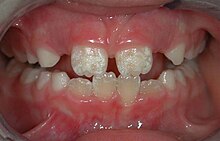
Irreversible enamel defects caused by an untreated celiac disease. They may be the only clue to its diagnosis, even in absence of gastrointestinal symptoms, but are often confused with fluorosis, tetracycline discoloration, acid reflux or other causes.[10][11][12] The National Institutes of Health include a dental exam in the diagnostic protocol of celiac disease.[10]

Dentists also encourage the prevention of oral diseases through proper hygiene and regular, twice or more yearly, checkups for professional cleaning and evaluation. Oral infections and inflammations may affect overall health and conditions in the oral cavity may be indicative of systemic diseases, such as osteoporosis, diabetes, celiac disease or cancer.[7][10][13][14] Many studies have also shown that gum disease is associated with an increased risk of diabetes, heart disease, and preterm birth. The concept that oral health can affect systemic health and disease is referred to as "oral-systemic health".

Education and licensing

[edit]
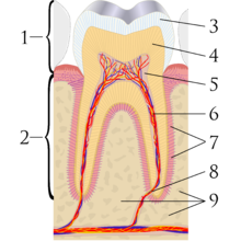
A sagittal cross-section of a molar tooth; 1: crown, 2: root, 3: enamel, 4: dentin and dentin tubules, 5: pulp chamber, 6: blood vessels and nerve, 7: periodontal ligament, 8: apex and periapical region, 9: alveolar bone
Early dental chair in Pioneer West Museum in Shamrock, Texas

John M. Harris started the world's first dental school in Bainbridge, Ohio, and helped to establish dentistry as a health profession. It opened on 21 February 1828, and today is a dental museum.[15] The first dental college, Baltimore College of Dental Surgery, opened in Baltimore, Maryland, US in 1840. The second in the United States was the Ohio College of Dental Surgery, established in Cincinnati, Ohio, in 1845.[16] The Philadelphia College of Dental Surgery followed in 1852.[17] In 1907, Temple University accepted a bid to incorporate the school.

Studies show that dentists that graduated from different countries,[18] or even from different dental schools in one country,[19] may make different clinical decisions for the same clinical condition. For example, dentists that graduated from Israeli dental schools may recommend the removal of asymptomatic impacted third molar (wisdom teeth) more often than dentists that graduated from Latin American or Eastern European dental schools.[20]

In the United Kingdom, the first dental schools, the London School of Dental Surgery and the Metropolitan School of Dental Science, both in London, opened in 1859.[21] The British Dentists Act of 1878 and the 1879 Dentists Register limited the title of "dentist" and "dental surgeon" to qualified and registered practitioners.[22][23] However, others could legally describe themselves as "dental experts" or "dental consultants".[24] The practice of dentistry in the United Kingdom became fully regulated with the 1921 Dentists Act, which required the registration of anyone practising dentistry.[25] The British Dental Association, formed in 1880 with Sir John Tomes as president, played a major role in prosecuting dentists practising illegally.[22] Dentists in the United Kingdom are now regulated by the General Dental Council.

In many countries, dentists usually complete between five and eight years of post-secondary education before practising. Though not mandatory, many dentists choose to complete an internship or residency focusing on specific aspects of dental care after they have received their dental degree. In a few countries, to become a qualified dentist one must usually complete at least four years of postgraduate study;[26] Dental degrees awarded around the world include the Doctor of Dental Surgery (DDS) and Doctor of Dental Medicine (DMD) in North America (US and Canada), and the Bachelor of Dental Surgery/Baccalaureus Dentalis Chirurgiae (BDS, BDent, BChD, BDSc) in the UK and current and former British Commonwealth countries.

All dentists in the United States undergo at least three years of undergraduate studies, but nearly all complete a bachelor's degree. This schooling is followed by four years of dental school to qualify as a "Doctor of Dental Surgery" (DDS) or "Doctor of Dental Medicine" (DMD). Specialization in dentistry is available in the fields of Anesthesiology, Dental Public Health, Endodontics, Oral Radiology, Oral and Maxillofacial Surgery, Oral Medicine, Orofacial Pain, Pathology, Orthodontics, Pediatric Dentistry (Pedodontics), Periodontics, and Prosthodontics.[27]

Specialties

[edit]
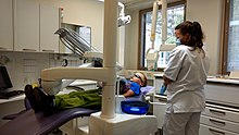
A modern dental clinic in Lappeenranta, Finland

Some dentists undertake further training after their initial degree in order to specialize. Exactly which subjects are recognized by dental registration bodies varies according to location. Examples include:

  • Anesthesiology[28] – The specialty of dentistry that deals with the advanced use of general anesthesia, sedation and pain management to facilitate dental procedures.
  • Cosmetic dentistry – Focuses on improving the appearance of the mouth, teeth and smile.
  • Dental public health – The study of epidemiology and social health policies relevant to oral health.
  • Endodontics (also called endodontology) – Root canal therapy and study of diseases of the dental pulp and periapical tissues.
  • Forensic odontology – The gathering and use of dental evidence in law. This may be performed by any dentist with experience or training in this field. The function of the forensic dentist is primarily documentation and verification of identity.
  • Geriatric dentistry or geriodontics – The delivery of dental care to older adults involving the diagnosis, prevention, and treatment of problems associated with normal aging and age-related diseases as part of an interdisciplinary team with other health care professionals.
  • Oral and maxillofacial pathology – The study, diagnosis, and sometimes the treatment of oral and maxillofacial related diseases.
  • Oral and maxillofacial radiology – The study and radiologic interpretation of oral and maxillofacial diseases.
  • Oral and maxillofacial surgery (also called oral surgery) – Extractions, implants, and surgery of the jaws, mouth and face.[nb 2]
  • Oral biology – Research in dental and craniofacial biology
  • Oral Implantology – The art and science of replacing extracted teeth with dental implants.
  • Oral medicine – The clinical evaluation and diagnosis of oral mucosal diseases
  • Orthodontics and dentofacial orthopedics – The straightening of teeth and modification of midface and mandibular growth.
  • Pediatric dentistry (also called pedodontics) – Dentistry for children
  • Periodontology (also called periodontics) – The study and treatment of diseases of the periodontium (non-surgical and surgical) as well as placement and maintenance of dental implants
  • Prosthodontics (also called prosthetic dentistry) – Dentures, bridges and the restoration of implants.
    • Some prosthodontists super-specialize in maxillofacial prosthetics, which is the discipline originally concerned with the rehabilitation of patients with congenital facial and oral defects such as cleft lip and palate or patients born with an underdeveloped ear (microtia). Today, most maxillofacial prosthodontists return function and esthetics to patients with acquired defects secondary to surgical removal of head and neck tumors, or secondary to trauma from war or motor vehicle accidents.
  • Special needs dentistry (also called special care dentistry) – Dentistry for those with developmental and acquired disabilities.
  • Sports dentistry – the branch of sports medicine dealing with prevention and treatment of dental injuries and oral diseases associated with sports and exercise.[29] The sports dentist works as an individual consultant or as a member of the Sports Medicine Team.
  • Veterinary dentistry – The field of dentistry applied to the care of animals. It is a specialty of veterinary medicine.[30][31]

History

[edit]
A wealthy patient falling over because of having a tooth extracted with such vigour by a fashionable dentist, c. 1790. History of Dentistry.
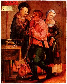
Farmer at the dentist, Johann Liss, c. 1616–17

Tooth decay was low in pre-agricultural societies, but the advent of farming society about 10,000 years ago correlated with an increase in tooth decay (cavities).[32] An infected tooth from Italy partially cleaned with flint tools, between 13,820 and 14,160 years old, represents the oldest known dentistry,[33] although a 2017 study suggests that 130,000 years ago the Neanderthals already used rudimentary dentistry tools.[34] In Italy evidence dated to the Paleolithic, around 13,000 years ago, points to bitumen used to fill a tooth[35] and in Neolithic Slovenia, 6500 years ago, beeswax was used to close a fracture in a tooth.[36] The Indus valley has yielded evidence of dentistry being practised as far back as 7000 BC, during the Stone Age.[37] The Neolithic site of Mehrgarh (now in Pakistan's south western province of Balochistan) indicates that this form of dentistry involved curing tooth related disorders with bow drills operated, perhaps, by skilled bead-crafters.[3] The reconstruction of this ancient form of dentistry showed that the methods used were reliable and effective.[38] The earliest dental filling, made of beeswax, was discovered in Slovenia and dates from 6500 years ago.[39] Dentistry was practised in prehistoric Malta, as evidenced by a skull which had a dental abscess lanced from the root of a tooth dating back to around 2500 BC.[40]

An ancient Sumerian text describes a "tooth worm" as the cause of dental caries.[41] Evidence of this belief has also been found in ancient India, Egypt, Japan, and China. The legend of the worm is also found in the Homeric Hymns,[42] and as late as the 14th century AD the surgeon Guy de Chauliac still promoted the belief that worms cause tooth decay.[43]

Recipes for the treatment of toothache, infections and loose teeth are spread throughout the Ebers Papyrus, Kahun Papyri, Brugsch Papyrus, and Hearst papyrus of Ancient Egypt.[44] The Edwin Smith Papyrus, written in the 17th century BC but which may reflect previous manuscripts from as early as 3000 BC, discusses the treatment of dislocated or fractured jaws.[44][45] In the 18th century BC, the Code of Hammurabi referenced dental extraction twice as it related to punishment.[46] Examination of the remains of some ancient Egyptians and Greco-Romans reveals early attempts at dental prosthetics.[47] However, it is possible the prosthetics were prepared after death for aesthetic reasons.[44]

Ancient Greek scholars Hippocrates and Aristotle wrote about dentistry, including the eruption pattern of teeth, treating decayed teeth and gum disease, extracting teeth with forceps, and using wires to stabilize loose teeth and fractured jaws.[48] Use of dental appliances, bridges and dentures was applied by the Etruscans in northern Italy, from as early as 700 BC, of human or other animal teeth fastened together with gold bands.[49][50][51] The Romans had likely borrowed this technique by the 5th century BC.[50][52] The Phoenicians crafted dentures during the 6th–4th century BC, fashioning them from gold wire and incorporating two ivory teeth.[53] In ancient Egypt, Hesy-Ra is the first named "dentist" (greatest of the teeth). The Egyptians bound replacement teeth together with gold wire. Roman medical writer Cornelius Celsus wrote extensively of oral diseases as well as dental treatments such as narcotic-containing emollients and astringents.[54] The earliest dental amalgams were first documented in a Tang dynasty medical text written by the Chinese physician Su Kung in 659, and appeared in Germany in 1528.[55][56]

During the Islamic Golden Age Dentistry was discussed in several famous books of medicine such as The Canon in medicine written by Avicenna and Al-Tasreef by Al-Zahrawi who is considered the greatest surgeon of the Middle Ages,[57] Avicenna said that jaw fracture should be reduced according to the occlusal guidance of the teeth; this principle is still valid in modern times. Al-Zahrawi invented over 200 surgical tools that resemble the modern kind.[58]

Historically, dental extractions have been used to treat a variety of illnesses. During the Middle Ages and throughout the 19th century, dentistry was not a profession in itself, and often dental procedures were performed by barbers or general physicians. Barbers usually limited their practice to extracting teeth which alleviated pain and associated chronic tooth infection. Instruments used for dental extractions date back several centuries. In the 14th century, Guy de Chauliac most probably invented the dental pelican[59] (resembling a pelican's beak) which was used to perform dental extractions up until the late 18th century. The pelican was replaced by the dental key[60] which, in turn, was replaced by modern forceps in the 19th century.[61]

Dental needle-nose pliers designed by Fauchard in the late 17th century to use in prosthodontics

The first book focused solely on dentistry was the "Artzney Buchlein" in 1530,[48] and the first dental textbook written in English was called "Operator for the Teeth" by Charles Allen in 1685.[23]

In the United Kingdom, there was no formal qualification for the providers of dental treatment until 1859 and it was only in 1921 that the practice of dentistry was limited to those who were professionally qualified. The Royal Commission on the National Health Service in 1979 reported that there were then more than twice as many registered dentists per 10,000 population in the UK than there were in 1921.[62]

Modern dentistry

[edit]
A microscopic device used in dental analysis, c. 1907

It was between 1650 and 1800 that the science of modern dentistry developed. The English physician Thomas Browne in his A Letter to a Friend (c. 1656 pub. 1690) made an early dental observation with characteristic humour:

The Egyptian Mummies that I have seen, have had their Mouths open, and somewhat gaping, which affordeth a good opportunity to view and observe their Teeth, wherein 'tis not easie to find any wanting or decayed: and therefore in Egypt, where one Man practised but one Operation, or the Diseases but of single Parts, it must needs be a barren Profession to confine unto that of drawing of Teeth, and little better than to have been Tooth-drawer unto King Pyrrhus, who had but two in his Head.

The French surgeon Pierre Fauchard became known as the "father of modern dentistry". Despite the limitations of the primitive surgical instruments during the late 17th and early 18th century, Fauchard was a highly skilled surgeon who made remarkable improvisations of dental instruments, often adapting tools from watchmakers, jewelers and even barbers, that he thought could be used in dentistry. He introduced dental fillings as treatment for dental cavities. He asserted that sugar-derived acids like tartaric acid were responsible for dental decay, and also suggested that tumors surrounding the teeth and in the gums could appear in the later stages of tooth decay.[63][64]

Panoramic radiograph of historic dental implants, made 1978

Fauchard was the pioneer of dental prosthesis, and he invented many methods to replace lost teeth. He suggested that substitutes could be made from carved blocks of ivory or bone. He also introduced dental braces, although they were initially made of gold, he discovered that the teeth position could be corrected as the teeth would follow the pattern of the wires. Waxed linen or silk threads were usually employed to fasten the braces. His contributions to the world of dental science consist primarily of his 1728 publication Le chirurgien dentiste or The Surgeon Dentist. The French text included "basic oral anatomy and function, dental construction, and various operative and restorative techniques, and effectively separated dentistry from the wider category of surgery".[63][64]

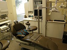
A modern dentist's chair

After Fauchard, the study of dentistry rapidly expanded. Two important books, Natural History of Human Teeth (1771) and Practical Treatise on the Diseases of the Teeth (1778), were published by British surgeon John Hunter. In 1763, he entered into a period of collaboration with the London-based dentist James Spence. He began to theorise about the possibility of tooth transplants from one person to another. He realised that the chances of a successful tooth transplant (initially, at least) would be improved if the donor tooth was as fresh as possible and was matched for size with the recipient. These principles are still used in the transplantation of internal organs. Hunter conducted a series of pioneering operations, in which he attempted a tooth transplant. Although the donated teeth never properly bonded with the recipients' gums, one of Hunter's patients stated that he had three which lasted for six years, a remarkable achievement for the period.[65]

Major advances in science were made in the 19th century, and dentistry evolved from a trade to a profession. The profession came under government regulation by the end of the 19th century. In the UK, the Dentist Act was passed in 1878 and the British Dental Association formed in 1879. In the same year, Francis Brodie Imlach was the first ever dentist to be elected President of the Royal College of Surgeons (Edinburgh), raising dentistry onto a par with clinical surgery for the first time.[66]

Hazards in modern dentistry

[edit]

Long term occupational noise exposure can contribute to permanent hearing loss, which is referred to as noise-induced hearing loss (NIHL) and tinnitus. Noise exposure can cause excessive stimulation of the hearing mechanism, which damages the delicate structures of the inner ear.[67] NIHL can occur when an individual is exposed to sound levels above 90 dBA according to the Occupational Safety and Health Administration (OSHA). Regulations state that the permissible noise exposure levels for individuals is 90 dBA.[68] For the National Institute for Occupational Safety and Health (NIOSH), exposure limits are set to 85 dBA. Exposures below 85 dBA are not considered to be hazardous. Time limits are placed on how long an individual can stay in an environment above 85 dBA before it causes hearing loss. OSHA places that limitation at 8 hours for 85 dBA. The exposure time becomes shorter as the dBA level increases.

Within the field of dentistry, a variety of cleaning tools are used including piezoelectric and sonic scalers, and ultrasonic scalers and cleaners.[69] While a majority of the tools do not exceed 75 dBA,[70] prolonged exposure over many years can lead to hearing loss or complaints of tinnitus.[71] Few dentists have reported using personal hearing protective devices,[72][73] which could offset any potential hearing loss or tinnitus.

Evidence-based dentistry

[edit]

There is a movement in modern dentistry to place a greater emphasis on high-quality scientific evidence in decision-making. Evidence-based dentistry (EBD) uses current scientific evidence to guide decisions. It is an approach to oral health that requires the application and examination of relevant scientific data related to the patient's oral and medical health. Along with the dentist's professional skill and expertise, EBD allows dentists to stay up to date on the latest procedures and patients to receive improved treatment. A new paradigm for medical education designed to incorporate current research into education and practice was developed to help practitioners provide the best care for their patients.[74] It was first introduced by Gordon Guyatt and the Evidence-Based Medicine Working Group at McMaster University in Ontario, Canada in the 1990s. It is part of the larger movement toward evidence-based medicine and other evidence-based practices, especially since a major part of dentistry involves dealing with oral and systemic diseases. Other issues relevant to the dental field in terms of evidence-based research and evidence-based practice include population oral health, dental clinical practice, tooth morphology etc.

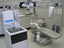
A dental chair at the University of Michigan School of Dentistry

Ethical and medicolegal issues

[edit]

Dentistry is unique in that it requires dental students to have competence-based clinical skills that can only be acquired through supervised specialized laboratory training and direct patient care.[75] This necessitates the need for a scientific and professional basis of care with a foundation of extensive research-based education.[76] According to some experts, the accreditation of dental schools can enhance the quality and professionalism of dental education.[77][78]

See also

[edit]
  • Dental aerosol
  • Dental instrument
  • Dental public health
  • Domestic healthcare:
    • Dentistry in ancient Rome
    • Dentistry in Canada
    • Dentistry in the Philippines
    • Dentistry in Israel
    • Dentistry in the United Kingdom
    • Dentistry in the United States
  • Eco-friendly dentistry
  • Geriatric dentistry
  • List of dental organizations
  • Pediatric dentistry
  • Sustainable dentistry
  • Veterinary dentistry
 

Notes

[edit]
  1. ^ Whether Dentists are referred to as "Doctor" is subject to geographic variation. For example, they are called "Doctor" in the US. In the UK, dentists have traditionally been referred to as "Mister" as they identified themselves with barber surgeons more than physicians (as do surgeons in the UK, see Surgeon#Titles). However more UK dentists now refer to themselves as "Doctor", although this was considered to be potentially misleading by the British public in a single report (see Costley and Fawcett 2010).
  2. ^ The scope of oral and maxillofacial surgery is variable. In some countries, both a medical and dental degree is required for training, and the scope includes head and neck oncology and craniofacial deformity.

References

[edit]
  1. ^ Neil Costley; Jo Fawcett (November 2010). General Dental Council Patient and Public Attitudes to Standards for Dental Professionals, Ethical Guidance and Use of the Term Doctor (PDF) (Report). General Dental Council/George Street Research. Archived from the original (PDF) on 4 March 2016. Retrieved 11 January 2017.
  2. ^ "Glossary of Dental Clinical and Administrative Terms". American Dental Association. Archived from the original on 6 March 2016. Retrieved 1 February 2014.
  3. ^ a b "Stone age man used dentist drill". BBC News. 6 April 2006. Retrieved 24 May 2010.
  4. ^ Suddick, RP; Harris, NO (1990). "Historical perspectives of oral biology: a series". Critical Reviews in Oral Biology and Medicine. 1 (2): 135–51. doi:10.1177/10454411900010020301. PMID 2129621.
  5. ^ "When barbers were surgeons and surgeons were barbers". Radio National. 15 April 2015. Retrieved 10 September 2021.
  6. ^ "dentistry". Etymonline.com. Retrieved 17 May 2018.
  7. ^ a b Gambhir RS (2015). "Primary care in dentistry – an untapped potential". Journal of Family Medicine and Primary Care (Review). 4 (1): 13–18. doi:10.4103/2249-4863.152239. PMC 4366984. PMID 25810982.
  8. ^ "What is the burden of oral disease?". WHO. Archived from the original on 30 June 2004. Retrieved 6 June 2017.
  9. ^ "American Academy of Cosmetic Dentistry | Dental CE Courses". aacd.com. Retrieved 21 October 2019.
  10. ^ a b c "Diagnosis of Celiac Disease". National Institute of Health (NIH). Archived from the original on 15 May 2017. Retrieved 6 June 2017.cite web: CS1 maint: bot: original URL status unknown (link)
  11. ^ Dental Enamel Defects and Celiac Disease (PDF) (Report). National Institute of Health (NIH). Archived from the original (PDF) on 5 March 2016.
  12. ^ Pastore L, Carroccio A, Compilato D, Panzarella V, Serpico R, Lo Muzio L (2008). "Oral manifestations of celiac disease". J Clin Gastroenterol (Review). 42 (3): 224–32. doi:10.1097/MCG.0b013e318074dd98. hdl:10447/1671. PMID 18223505. S2CID 205776755.
  13. ^ Estrella MR, Boynton JR (2010). "General dentistry's role in the care for children with special needs: a review". Gen Dent (Review). 58 (3): 222–29. PMID 20478802.
  14. ^ da Fonseca MA (2010). "Dental and oral care for chronically ill children and adolescents". Gen Dent (Review). 58 (3): 204–09, quiz 210–11. PMID 20478800.
  15. ^ Owen, Lorrie K., ed. (1999). Dictionary of Ohio Historic Places. Vol. 2. St. Clair Shores: Somerset. pp. 1217–1218.
  16. ^ Mary, Otto (2017). Teeth: the story of beauty, inequality, and the struggle for oral health in America. New York: The New Press. p. 70. ISBN 978-1-62097-144-4. OCLC 958458166.
  17. ^ "History". Pennsylvania School of Dental Medicine. Retrieved 13 January 2016.
  18. ^ Zadik Yehuda; Levin Liran (January 2008). "Clinical decision making in restorative dentistry, endodontics, and antibiotic prescription". J Dent Educ. 72 (1): 81–86. doi:10.1002/j.0022-0337.2008.72.1.tb04456.x. PMID 18172239.
  19. ^ Zadik Yehuda; Levin Liran (April 2006). "Decision making of Hebrew University and Tel Aviv University Dental Schools graduates in every day dentistry—is there a difference?". J Isr Dent Assoc. 23 (2): 19–23. PMID 16886872.
  20. ^ Zadik Yehuda; Levin Liran (April 2007). "Decision making of Israeli, East European, and South American dental school graduates in third molar surgery: is there a difference?". J Oral Maxillofac Surg. 65 (4): 658–62. doi:10.1016/j.joms.2006.09.002. PMID 17368360.
  21. ^ Gelbier, Stanley (1 October 2005). "Dentistry and the University of London". Medical History. 49 (4): 445–462. doi:10.1017/s0025727300009157. PMC 1251639. PMID 16562330.
  22. ^ a b Gelbier, S. (2005). "125 years of developments in dentistry, 1880–2005 Part 2: Law and the dental profession". British Dental Journal. 199 (7): 470–473. doi:10.1038/sj.bdj.4812875. ISSN 1476-5373. PMID 16215593. The 1879 register is referred to as the "Dental Register".
  23. ^ a b "The story of dentistry: Dental History Timeline". British Dental Association. Archived from the original on 9 March 2012. Retrieved 2 March 2010.
  24. ^ J Menzies Campbell (8 February 1955). "Banning Clerks, Colliers and other Charlatans". The Glasgow Herald. p. 3. Retrieved 5 April 2017.
  25. ^ "History of Dental Surgery in Edinburgh" (PDF). Royal College of Surgeons of Edinburgh. Retrieved 11 December 2007.
  26. ^ "Dentistry (D.D.S. or D.M.D.)" (PDF). Purdue.edu. Archived from the original (PDF) on 9 January 2017. Retrieved 17 May 2018.
  27. ^ "Canadian Dental Association". cda-adc.ca. Retrieved 21 October 2019.
  28. ^ "Anesthesiology recognized as a dental specialty". www.ada.org. Archived from the original on 21 September 2019. Retrieved 12 March 2019.
  29. ^ "Sports dentistry". FDI World Dental Federation. Archived from the original on 23 October 2020. Retrieved 13 July 2020.
  30. ^ "AVDC Home". Avdc.org. 29 November 2009. Retrieved 18 April 2010.
  31. ^ "EVDC web site". Evdc.info. Archived from the original on 5 September 2018. Retrieved 18 April 2010.
  32. ^ Barras, Colin (29 February 2016). "How our ancestors drilled rotten teeth". BBC. Archived from the original on 19 May 2017. Retrieved 1 March 2016.
  33. ^ "Oldest Dentistry Found in 14,000-Year-Old Tooth". Discovery Channel. 16 July 2015. Archived from the original on 18 July 2015. Retrieved 21 July 2015.
  34. ^ "Analysis of Neanderthal teeth marks uncovers evidence of prehistoric dentistry". The University of Kansas. 28 June 2017. Retrieved 1 July 2017.
  35. ^ Oxilia, Gregorio; Fiorillo, Flavia; Boschin, Francesco; Boaretto, Elisabetta; Apicella, Salvatore A.; Matteucci, Chiara; Panetta, Daniele; Pistocchi, Rossella; Guerrini, Franca; Margherita, Cristiana; Andretta, Massimo; Sorrentino, Rita; Boschian, Giovanni; Arrighi, Simona; Dori, Irene (2017). "The dawn of dentistry in the late upper Paleolithic: An early case of pathological intervention at Riparo Fredian". American Journal of Physical Anthropology. 163 (3): 446–461. doi:10.1002/ajpa.23216. hdl:11585/600517. ISSN 0002-9483. PMID 28345756.
  36. ^ Bernardini, Federico; Tuniz, Claudio; Coppa, Alfredo; Mancini, Lucia; Dreossi, Diego; Eichert, Diane; Turco, Gianluca; Biasotto, Matteo; Terrasi, Filippo; Cesare, Nicola De; Hua, Quan; Levchenko, Vladimir (19 September 2012). "Beeswax as Dental Filling on a Neolithic Human Tooth". PLOS ONE. 7 (9): e44904. Bibcode:2012PLoSO...744904B. doi:10.1371/journal.pone.0044904. ISSN 1932-6203. PMC 3446997. PMID 23028670.
  37. ^ Coppa, A.; et al. (2006). "Early Neolithic tradition of dentistry". Nature. 440 (7085). Springer Science and Business Media LLC: 755–756. doi:10.1038/440755a. ISSN 0028-0836. PMID 16598247.
  38. ^ "Dig uncovers ancient roots of dentistry". NBC News. 5 April 2006.
  39. ^ Bernardini, Federico; et al. (2012). "Beeswax as Dental Filling on a Neolithic Human Tooth". PLOS ONE. 7 (9): e44904. Bibcode:2012PLoSO...744904B. doi:10.1371/journal.pone.0044904. PMC 3446997. PMID 23028670.
  40. ^ "700 years added to Malta's history". Times of Malta. 16 March 2018. Archived from the original on 16 March 2018.
  41. ^ "History of Dentistry: Ancient Origins". American Dental Association. Archived from the original on 5 July 2007. Retrieved 9 January 2007.
  42. ^ TOWNEND, B. R. (1944). "The Story of the Tooth-Worm". Bulletin of the History of Medicine. 15 (1): 37–58. ISSN 0007-5140. JSTOR 44442797.
  43. ^ Suddick Richard P., Harris Norman O. (1990). "Historical Perspectives of Oral Biology: A Series" (PDF). Critical Reviews in Oral Biology and Medicine. 1 (2): 135–51. doi:10.1177/10454411900010020301. PMID 2129621. Archived from the original (PDF) on 18 December 2007.
  44. ^ a b c Blomstedt, P. (2013). "Dental surgery in ancient Egypt". Journal of the History of Dentistry. 61 (3): 129–42. PMID 24665522.
  45. ^ "Ancient Egyptian Dentistry". University of Oklahoma. Archived from the original on 26 December 2007. Retrieved 15 December 2007.
  46. ^ Wilwerding, Terry. "History of Dentistry 2001" (PDF). Archived from the original (PDF) on 3 November 2014. Retrieved 3 November 2014.
  47. ^ "Medicine in Ancient Egypt 3". Arabworldbooks.com. Retrieved 18 April 2010.
  48. ^ a b "History Of Dentistry". Complete Dental Guide. Archived from the original on 14 July 2016. Retrieved 29 June 2016.
  49. ^ "History of Dentistry Research Page, Newsletter". Rcpsg.ac.uk. Archived from the original on 28 April 2015. Retrieved 9 June 2014.
  50. ^ a b Donaldson, J. A. (1980). "The use of gold in dentistry" (PDF). Gold Bulletin. 13 (3): 117–124. doi:10.1007/BF03216551. PMID 11614516. S2CID 137571298.
  51. ^ Becker, Marshall J. (1999). Ancient "dental implants": a recently proposed example from France evaluated with other spurious examples (PDF). International Journal of Oral & Maxillofacial Implants 14.1.
  52. ^ Malik, Ursman. "History of Dentures from Beginning to Early 19th Century". Exhibits. Retrieved 3 May 2023.
  53. ^ Renfrew, Colin; Bahn, Paul (2012). Archaeology: Theories, Methods, and Practice (6th ed.). Thames & Hudson. p. 449. ISBN 978-0-500-28976-1.
  54. ^ "Dental Treatment in the Ancient Times". Dentaltreatment.org.uk. Archived from the original on 1 December 2009. Retrieved 18 April 2010.
  55. ^ Bjørklund G (1989). "The history of dental amalgam (in Norwegian)". Tidsskr Nor Laegeforen. 109 (34–36): 3582–85. PMID 2694433.
  56. ^ Czarnetzki, A.; Ehrhardt S. (1990). "Re-dating the Chinese amalgam-filling of teeth in Europe". International Journal of Anthropology. 5 (4): 325–32.
  57. ^ Meri, Josef (2005). Medieval Islamic Civilization: An Encyclopedia (Routledge Encyclopedias of the Middle Ages). Psychology Press. ISBN 978-0-415-96690-0.
  58. ^ Friedman, Saul S. (2006). A history of the Middle East. Jefferson, N.C.: Mcfarland. p. 152. ISBN 0786451343.
  59. ^ Gregory Ribitzky. "Pelican". Archived from the original on 25 January 2020. Retrieved 23 June 2018.
  60. ^ Gregory Ribitzky. "Toothkey". Archived from the original on 23 June 2018. Retrieved 23 June 2018.
  61. ^ Gregory Ribitzky. "Forceps". Archived from the original on 23 June 2018. Retrieved 23 June 2018.
  62. ^ Royal Commission on the NHS Chapter 9. HMSO. July 1979. ISBN 978-0-10-176150-5. Retrieved 19 May 2015.
  63. ^ a b André Besombes; Phillipe de Gaillande (1993). Pierre Fauchard (1678–1761): The First Dental Surgeon, His Work, His Actuality. Pierre Fauchard Academy.
  64. ^ a b Bernhard Wolf Weinberger (1941). Pierre Fauchard, Surgeon-dentist: A Brief Account of the Beginning of Modern Dentistry, the First Dental Textbook, and Professional Life Two Hundred Years Ago. Pierre Fauchard Academy.
  65. ^ Moore, Wendy (30 September 2010). The Knife Man. Transworld. pp. 223–24. ISBN 978-1-4090-4462-8. Retrieved 8 March 2012.
  66. ^ Dingwall, Helen (April 2004). "A pioneering history: dentistry and the Royal College of Surgeons of Edinburgh" (PDF). History of Dentistry Newsletter. No. 14. Archived from the original (PDF) on 1 February 2013.
  67. ^ "Noise-Induced Hearing Loss". NIDCD. 18 August 2015.
  68. ^ "Occupational Safety and Health Standards | Occupational Safety and Health Administration". Osha.gov.
  69. ^ Stevens, M (1999). "Is someone listening to the din of occupational noise exposure in dentistry". RDH (19): 34–85.
  70. ^ Merrel, HB (1992). "Noise pollution and hearing loss in the dental office". Dental Assisting Journal. 61 (3): 6–9.
  71. ^ Wilson, J.D. (2002). "Effects of occupational ultrasonic noise exposure on hearing of dental hygienists: A pilot study". Journal of Dental Hygiene. 76 (4): 262–69. PMID 12592917.
  72. ^ Leggat, P.A. (2007). "Occupational Health Problems in Modern Dentistry: A Review" (PDF). Industrial Health. 45 (5): 611–21. doi:10.2486/indhealth.45.611. PMID 18057804. Archived (PDF) from the original on 27 April 2019.
  73. ^ Leggat, P.A. (2001). "Occupational hygiene practices of dentists in southern Thailand". International Dental Journal. 51 (51): 11–6. doi:10.1002/j.1875-595x.2001.tb00811.x. PMID 11326443.
  74. ^ Evidence-Based Medicine Working Group (1992). "Evidence-based medicine. A new approach to teaching the practice of medicine". Journal of the American Medical Association. 268 (17): 2420–2425. doi:10.1001/jama.1992.03490170092032. PMID 1404801.
  75. ^ "Union workers build high-tech dental simulation laboratory for SIU dental school". The Labor Tribune. 17 March 2014. Retrieved 10 September 2021.
  76. ^ Slavkin, Harold C. (January 2012). "Evolution of the scientific basis for dentistry and its impact on dental education: past, present, and future". Journal of Dental Education. 76 (1): 28–35. doi:10.1002/j.0022-0337.2012.76.1.tb05231.x. ISSN 1930-7837. PMID 22262547.
  77. ^ Formicola, Allan J.; Bailit, Howard L.; Beazoglou, Tryfon J.; Tedesco, Lisa A. (February 2008). "The interrelationship of accreditation and dental education: history and current environment". Journal of Dental Education. 72 (2 Suppl): 53–60. doi:10.1002/j.0022-0337.2008.72.2_suppl.tb04480.x. ISSN 0022-0337. PMID 18250379.
  78. ^ Carrrassi, A. (2019). "The first 25 year [Internet] Ireland: ADEE (Association for Dental Education in Europe)". Association for Dental Education in Europe. Retrieved 21 October 2019.
[edit]


Tooth
A chimpanzee displaying his teeth
Details
Identifiers
Latin dens
MeSH D014070
FMA 12516
Anatomical terminology
[edit on Wikidata]

A tooth (pl.: teeth) is a hard, calcified structure found in the jaws (or mouths) of many vertebrates and used to break down food. Some animals, particularly carnivores and omnivores, also use teeth to help with capturing or wounding prey, tearing food, for defensive purposes, to intimidate other animals often including their own, or to carry prey or their young. The roots of teeth are covered by gums. Teeth are not made of bone, but rather of multiple tissues of varying density and hardness that originate from the outermost embryonic germ layer, the ectoderm.

The general structure of teeth is similar across the vertebrates, although there is considerable variation in their form and position. The teeth of mammals have deep roots, and this pattern is also found in some fish, and in crocodilians. In most teleost fish, however, the teeth are attached to the outer surface of the bone, while in lizards they are attached to the inner surface of the jaw by one side. In cartilaginous fish, such as sharks, the teeth are attached by tough ligaments to the hoops of cartilage that form the jaw.[1]

Monophyodonts are animals that develop only one set of teeth, while diphyodonts grow an early set of deciduous teeth and a later set of permanent or "adult" teeth. Polyphyodonts grow many sets of teeth. For example, sharks, grow a new set of teeth every two weeks to replace worn teeth. Most extant mammals including humans are diphyodonts, but there are exceptions including elephants, kangaroos, and manatees, all of which are polyphyodonts.

Rodent incisors grow and wear away continually through gnawing, which helps maintain relatively constant length. The industry of the beaver is due in part to this qualification. Some rodents, such as voles and guinea pigs (but not mice), as well as lagomorpha (rabbits, hares and pikas), have continuously growing molars in addition to incisors.[2][3] Also, tusks (in tusked mammals) grow almost throughout life.[4]

Teeth are not always attached to the jaw, as they are in mammals. In many reptiles and fish, teeth are attached to the palate or to the floor of the mouth, forming additional rows inside those on the jaws proper. Some teleosts even have teeth in the pharynx. While not true teeth in the usual sense, the dermal denticles of sharks are almost identical in structure and are likely to have the same evolutionary origin. Indeed, teeth appear to have first evolved in sharks, and are not found in the more primitive jawless fish – while lampreys do have tooth-like structures on the tongue, these are in fact, composed of keratin, not of dentine or enamel, and bear no relationship to true teeth.[1] Though "modern" teeth-like structures with dentine and enamel have been found in late conodonts, they are now supposed to have evolved independently of later vertebrates' teeth.[5][6]

Living amphibians typically have small teeth, or none at all, since they commonly feed only on soft foods. In reptiles, teeth are generally simple and conical in shape, although there is some variation between species, most notably the venom-injecting fangs of snakes. The pattern of incisors, canines, premolars and molars is found only in mammals, and to varying extents, in their evolutionary ancestors. The numbers of these types of teeth vary greatly between species; zoologists use a standardised dental formula to describe the precise pattern in any given group.[1]

Etymology

[edit]

The word tooth comes from Proto-Germanic *tanþs, derived from the Proto-Indo-European *h₁dent-, which was composed of the root *h₁ed- 'to eat' plus the active participle suffix *-nt, therefore literally meaning 'that which eats'.[7]

The irregular plural form teeth is the result of Germanic umlaut whereby vowels immediately preceding a high vocalic in the following syllable were raised. As the nominative plural ending of the Proto-Germanic consonant stems (to which *tanþs belonged) was *-iz, the root vowel in the plural form *tanþiz (changed by this point to *tÄ…Ì„þi via unrelated phonological processes) was raised to /œÃ‹Â/, and later unrounded to /eː/, resulting in the tōþ/tÄ“þ alternation attested from Old English. Cf. also Old English bōc/bÄ“Ä‹ 'book/books' and 'mÅ«s/mȳs' 'mouse/mice', from Proto-Germanic *bōks/bōkiz and *mÅ«s/mÅ«siz respectively.

Cognate with Latin dÄ“ns, Greek á½€δούς (odous), and Sanskrit dát.

Origin

[edit]

Teeth are assumed to have evolved either from ectoderm denticles (scales, much like those on the skin of sharks) that folded and integrated into the mouth (called the "outside–in" theory), or from endoderm pharyngeal teeth (primarily formed in the pharynx of jawless vertebrates) (the "inside–out" theory). In addition, there is another theory stating that neural crest gene regulatory network, and neural crest-derived ectomesenchyme are the key to generate teeth (with any epithelium, either ectoderm or endoderm).[4][8]

The genes governing tooth development in mammals are homologous to those involved in the development of fish scales.[9] Study of a tooth plate of a fossil of the extinct fish Romundina stellina showed that the teeth and scales were made of the same tissues, also found in mammal teeth, lending support to the theory that teeth evolved as a modification of scales.[10]

Mammals

[edit]

Teeth are among the most distinctive (and long-lasting) features of mammal species. Paleontologists use teeth to identify fossil species and determine their relationships. The shape of the animal's teeth are related to its diet. For example, plant matter is hard to digest, so herbivores have many molars for chewing and grinding. Carnivores, on the other hand, have canine teeth to kill prey and to tear meat.

Mammals, in general, are diphyodont, meaning that they develop two sets of teeth. In humans, the first set (the "baby", "milk", "primary" or "deciduous" set) normally starts to appear at about six months of age, although some babies are born with one or more visible teeth, known as neonatal teeth. Normal tooth eruption at about six months is known as teething and can be painful. Kangaroos, elephants, and manatees are unusual among mammals because they are polyphyodonts.

Aardvark

[edit]

In aardvarks, teeth lack enamel and have many pulp tubules, hence the name of the order Tubulidentata.[11]

Canines

[edit]

In dogs, the teeth are less likely than humans to form dental cavities because of the very high pH of dog saliva, which prevents enamel from demineralizing.[12] Sometimes called cuspids, these teeth are shaped like points (cusps) and are used for tearing and grasping food.[13]

Cetaceans

[edit]

Like human teeth, whale teeth have polyp-like protrusions located on the root surface of the tooth. These polyps are made of cementum in both species, but in human teeth, the protrusions are located on the outside of the root, while in whales the nodule is located on the inside of the pulp chamber. While the roots of human teeth are made of cementum on the outer surface, whales have cementum on the entire surface of the tooth with a very small layer of enamel at the tip. This small enamel layer is only seen in older whales where the cementum has been worn away to show the underlying enamel.[14]

The toothed whale is a parvorder of the cetaceans characterized by having teeth. The teeth differ considerably among the species. They may be numerous, with some dolphins bearing over 100 teeth in their jaws. On the other hand, the narwhals have a giant unicorn-like tusk, which is a tooth containing millions of sensory pathways and used for sensing during feeding, navigation, and mating. It is the most neurologically complex tooth known. Beaked whales are almost toothless, with only bizarre teeth found in males. These teeth may be used for feeding but also for demonstrating aggression and showmanship.

Primates

[edit]

In humans (and most other primates), there are usually 20 primary (also "baby" or "milk") teeth, and later up to 32 permanent teeth. Four of these 32 may be third molars or wisdom teeth, although these are not present in all adults, and may be removed surgically later in life.[15]

Among primary teeth, 10 of them are usually found in the maxilla (i.e. upper jaw) and the other 10 in the mandible (i.e. lower jaw). Among permanent teeth, 16 are found in the maxilla and the other 16 in the mandible. Most of the teeth have uniquely distinguishing features.

Horse

[edit]

An adult horse has between 36 and 44 teeth. The enamel and dentin layers of horse teeth are intertwined.[16] All horses have 12 premolars, 12 molars, and 12 incisors.[17] Generally, all male equines also have four canine teeth (called tushes) between the molars and incisors. However, few female horses (less than 28%) have canines, and those that do usually have only one or two, which many times are only partially erupted.[18] A few horses have one to four wolf teeth, which are vestigial premolars, with most of those having only one or two. They are equally common in male and female horses and much more likely to be on the upper jaw. If present these can cause problems as they can interfere with the horse's bit contact. Therefore, wolf teeth are commonly removed.[17]

Horse teeth can be used to estimate the animal's age. Between birth and five years, age can be closely estimated by observing the eruption pattern on milk teeth and then permanent teeth. By age five, all permanent teeth have usually erupted. The horse is then said to have a "full" mouth. After the age of five, age can only be conjectured by studying the wear patterns on the incisors, shape, the angle at which the incisors meet, and other factors. The wear of teeth may also be affected by diet, natural abnormalities, and cribbing. Two horses of the same age may have different wear patterns.

A horse's incisors, premolars, and molars, once fully developed, continue to erupt as the grinding surface is worn down through chewing. A young adult horse will have teeth, which are 110–130 mm (4.5–5 inches) long, with the majority of the crown remaining below the gumline in the dental socket. The rest of the tooth will slowly emerge from the jaw, erupting about 3 mm (18 in) each year, as the horse ages. When the animal reaches old age, the crowns of the teeth are very short and the teeth are often lost altogether. Very old horses, if lacking molars, may need to have their fodder ground up and soaked in water to create a soft mush for them to eat in order to obtain adequate nutrition.

Proboscideans

[edit]
Section through the ivory tusk of a mammoth

Elephants' tusks are specialized incisors for digging food up and fighting. Some elephant teeth are similar to those in manatees, and elephants are believed to have undergone an aquatic phase in their evolution.

At birth, elephants have a total of 28 molar plate-like grinding teeth not including the tusks. These are organized into four sets of seven successively larger teeth which the elephant will slowly wear through during its lifetime of chewing rough plant material. Only four teeth are used for chewing at a given time, and as each tooth wears out, another tooth moves forward to take its place in a process similar to a conveyor belt. The last and largest of these teeth usually becomes exposed when the animal is around 40 years of age, and will often last for an additional 20 years. When the last of these teeth has fallen out, regardless of the elephant's age, the animal will no longer be able to chew food and will die of starvation.[19][20]

Rabbit

[edit]

Rabbits and other lagomorphs usually shed their deciduous teeth before (or very shortly after) their birth, and are usually born with their permanent teeth.[21] The teeth of rabbits complement their diet, which consists of a wide range of vegetation. Since many of the foods are abrasive enough to cause attrition, rabbit teeth grow continuously throughout life.[22] Rabbits have a total of six incisors, three upper premolars, three upper molars, two lower premolars, and two lower molars on each side. There are no canines. Dental formula is 2.0.3.31.0.2.3 = 28. Three to four millimeters of the tooth is worn away by incisors every week, whereas the cheek teeth require a month to wear away the same amount.[23]

The incisors and cheek teeth of rabbits are called aradicular hypsodont teeth. This is sometimes referred to as an elodent dentition. These teeth grow or erupt continuously. The growth or eruption is held in balance by dental abrasion from chewing a diet high in fiber.

Buccal view of top incisor from Rattus rattus. Top incisor outlined in yellow. Molars circled in blue.
Buccal view of the lower incisor from the right dentary of a Rattus rattus
Lingual view of the lower incisor from the right dentary of a Rattus rattus
Midsagittal view of top incisor from Rattus rattus. Top incisor outlined in yellow. Molars circled in blue.

Rodents

[edit]

Rodents have upper and lower hypselodont incisors that can continuously grow enamel throughout its life without having properly formed roots.[24] These teeth are also known as aradicular teeth, and unlike humans whose ameloblasts die after tooth development, rodents continually produce enamel, they must wear down their teeth by gnawing on various materials.[25] Enamel and dentin are produced by the enamel organ, and growth is dependent on the presence of stem cells, cellular amplification, and cellular maturation structures in the odontogenic region.[26] Rodent incisors are used for cutting wood, biting through the skin of fruit, or for defense. This allows for the rate of wear and tooth growth to be at equilibrium.[24] The microstructure of rodent incisor enamel has shown to be useful in studying the phylogeny and systematics of rodents because of its independent evolution from the other dental traits. The enamel on rodent incisors are composed of two layers: the inner portio interna (PI) with Hunter-Schreger bands (HSB) and an outer portio externa (PE) with radial enamel (RE).[27] It usually involves the differential regulation of the epithelial stem cell niche in the tooth of two rodent species, such as guinea pigs.[28][29]

Lingual view of top incisor from Rattus rattus. Top incisor outlined in yellow. Molars circled in blue.

The teeth have enamel on the outside and exposed dentin on the inside, so they self-sharpen during gnawing. On the other hand, continually growing molars are found in some rodent species, such as the sibling vole and the guinea pig.[28][29] There is variation in the dentition of the rodents, but generally, rodents lack canines and premolars, and have a space between their incisors and molars, called the diastema region.

Manatee

[edit]

Manatees are polyphyodont with mandibular molars developing separately from the jaw and are encased in a bony shell separated by soft tissue.[30][31]

Walrus

[edit]

Walrus tusks are canine teeth that grow continuously throughout life.[32]

Fish

[edit]
Teeth of a great white shark

Fish, such as sharks, may go through many teeth in their lifetime. The replacement of multiple teeth is known as polyphyodontia.

A class of prehistoric shark are called cladodonts for their strange forked teeth.

Unlike the continuous shedding of functional teeth seen in modern sharks,[33][34] the majority of stem chondrichthyan lineages retained all tooth generations developed throughout the life of the animal.[35] This replacement mechanism is exemplified by the tooth whorl-based dentitions of acanthodians,[36] which include the oldest known toothed vertebrate, Qianodus duplicis[37].

Amphibians

[edit]

All amphibians have pedicellate teeth, which are modified to be flexible due to connective tissue and uncalcified dentine that separates the crown from the base of the tooth.[38]

Most amphibians exhibit teeth that have a slight attachment to the jaw or acrodont teeth. Acrodont teeth exhibit limited connection to the dentary and have little enervation.[39] This is ideal for organisms who mostly use their teeth for grasping, but not for crushing and allows for rapid regeneration of teeth at a low energy cost. Teeth are usually lost in the course of feeding if the prey is struggling. Additionally, amphibians that undergo a metamorphosis develop bicuspid shaped teeth.[40]

Reptiles

[edit]

The teeth of reptiles are replaced constantly throughout their lives. Crocodilian juveniles replace teeth with larger ones at a rate as high as one new tooth per socket every month. Once mature, tooth replacement rates can slow to two years and even longer. Overall, crocodilians may use 3,000 teeth from birth to death. New teeth are created within old teeth.[41]

Birds

[edit]

A skull of Ichthyornis discovered in 2014 suggests that the beak of birds may have evolved from teeth to allow chicks to escape their shells earlier, and thus avoid predators and also to penetrate protective covers such as hard earth to access underlying food.[42][43]

Invertebrates

[edit]
The European medicinal leech has three jaws with numerous sharp teeth which function like little saws for incising a host.

True teeth are unique to vertebrates,[44] although many invertebrates have analogous structures often referred to as teeth. The organisms with the simplest genome bearing such tooth-like structures are perhaps the parasitic worms of the family Ancylostomatidae.[45] For example, the hookworm Necator americanus has two dorsal and two ventral cutting plates or teeth around the anterior margin of the buccal capsule. It also has a pair of subdorsal and a pair of subventral teeth located close to the rear.[46]

Historically, the European medicinal leech, another invertebrate parasite, has been used in medicine to remove blood from patients.[47] They have three jaws (tripartite) that resemble saws in both appearance and function, and on them are about 100 sharp teeth used to incise the host. The incision leaves a mark that is an inverted Y inside of a circle. After piercing the skin and injecting anticoagulants (hirudin) and anaesthetics, they suck out blood, consuming up to ten times their body weight in a single meal.[48]

In some species of Bryozoa, the first part of the stomach forms a muscular gizzard lined with chitinous teeth that crush armoured prey such as diatoms. Wave-like peristaltic contractions then move the food through the stomach for digestion.[49]

The limpet rasps algae from rocks using teeth with the strongest known tensile strength of any biological material.

Molluscs have a structure called a radula, which bears a ribbon of chitinous teeth. However, these teeth are histologically and developmentally different from vertebrate teeth and are unlikely to be homologous. For example, vertebrate teeth develop from a neural crest mesenchyme-derived dental papilla, and the neural crest is specific to vertebrates, as are tissues such as enamel.[44]

The radula is used by molluscs for feeding and is sometimes compared rather inaccurately to a tongue. It is a minutely toothed, chitinous ribbon, typically used for scraping or cutting food before the food enters the oesophagus. The radula is unique to molluscs, and is found in every class of mollusc apart from bivalves.

Within the gastropods, the radula is used in feeding by both herbivorous and carnivorous snails and slugs. The arrangement of teeth (also known as denticles) on the radula ribbon varies considerably from one group to another as shown in the diagram on the left.

Predatory marine snails such as the Naticidae use the radula plus an acidic secretion to bore through the shell of other molluscs. Other predatory marine snails, such as the Conidae, use a specialized radula tooth as a poisoned harpoon. Predatory pulmonate land slugs, such as the ghost slug, use elongated razor-sharp teeth on the radula to seize and devour earthworms. Predatory cephalopods, such as squid, use the radula for cutting prey.

In most of the more ancient lineages of gastropods, the radula is used to graze by scraping diatoms and other microscopic algae off rock surfaces and other substrates. Limpets scrape algae from rocks using radula equipped with exceptionally hard rasping teeth.[50] These teeth have the strongest known tensile strength of any biological material, outperforming spider silk.[50] The mineral protein of the limpet teeth can withstand a tensile stress of 4.9 GPa, compared to 4 GPa of spider silk and 0.5 GPa of human teeth.[51]


Fossilization and taphonomy

[edit]

Because teeth are very resistant, often preserved when bones are not,[52] and reflect the diet of the host organism, they are very valuable to archaeologists and palaeontologists.[53] Early fish such as the thelodonts had scales composed of dentine and an enamel-like compound, suggesting that the origin of teeth was from scales which were retained in the mouth. Fish as early as the late Cambrian had dentine in their exoskeletons, which may have functioned in defense or for sensing their environments.[54] Dentine can be as hard as the rest of teeth and is composed of collagen fibres, reinforced with hydroxyapatite.[54]

Though teeth are very resistant, they also can be brittle and highly susceptible to cracking.[55] However, cracking of the tooth can be used as a diagnostic tool for predicting bite force. Additionally, enamel fractures can also give valuable insight into the diet and behaviour of archaeological and fossil samples.

Decalcification removes the enamel from teeth and leaves only the organic interior intact, which comprises dentine and cementine.[56] Enamel is quickly decalcified in acids,[57] perhaps by dissolution by plant acids or via diagenetic solutions, or in the stomachs of vertebrate predators.[56] Enamel can be lost by abrasion or spalling,[56] and is lost before dentine or bone are destroyed by the fossilisation process.[57] In such a case, the 'skeleton' of the teeth would consist of the dentine, with a hollow pulp cavity.[56] The organic part of dentine, conversely, is destroyed by alkalis.[57]

See also

[edit]
  • Animal tooth development
  • Dragon's teeth (mythology)

References

[edit]
  1. ^ a b c Romer, Alfred Sherwood; Parsons, Thomas S. (1977). The Vertebrate Body. Philadelphia, PA: Holt-Saunders International. pp. 300–310. ISBN 978-0-03-910284-5.
  2. ^ Tummers M, Thesleff I (March 2003). "Root or crown: a developmental choice orchestrated by the differential regulation of the epithelial stem cell niche in the tooth of two rodent species". Development. 130 (6): 1049–57. doi:10.1242/dev.00332. PMID 12571097.
  3. ^ Hunt AM (1959). "A description of the molar teeth and investing tissues of normal guinea pigs". J. Dent. Res. 38 (2): 216–31. doi:10.1177/00220345590380020301. PMID 13641521. S2CID 45097018.
  4. ^ a b Nasoori, Alireza (2020). "Tusks, the extra-oral teeth". Archives of Oral Biology. 117: 104835. doi:10.1016/j.archoralbio.2020.104835. PMID 32668361. S2CID 220585014.
  5. ^ McCOLLUM, MELANIE; SHARPE, PAUL T. (July 2001). "Evolution and development of teeth". Journal of Anatomy. 199 (1–2): 153–159. doi:10.1046/j.1469-7580.2001.19910153.x. PMC 1594990. PMID 11523817.
  6. ^ Kaplan, Matt (October 16, 2013). "Fossil scans reveal origins of teeth". Nature. doi:10.1038/nature.2013.13964 – via www.nature.com.
  7. ^ Harper, Douglas (2001–2021). "tooth | Origin and meaning of tooth". Online Etymology Dictionary.
  8. ^ Jheon, Andrew H (2012). "From molecules to mastication: the development and evolution of teeth". Wiley Interdiscip Rev Dev Biol. 2 (2): 165–182. doi:10.1002/wdev.63. PMC 3632217. PMID 24009032.
  9. ^ Sharpe, P. T. (2001). "Fish scale development: Hair today, teeth and scales yesterday?". Current Biology. 11 (18): R751 – R752. Bibcode:2001CBio...11.R751S. doi:10.1016/S0960-9822(01)00438-9. PMID 11566120. S2CID 18868124.
  10. ^ Jennifer Viegas (June 24, 2015). "First-known teeth belonged to fierce fish". ABC Science. Retrieved June 28, 2015.
  11. ^ Shoshani 2002, p. 619
  12. ^ Hale, FA (2009). "Dental caries in the dog". Can. Vet. J. 50 (12): 1301–4. PMC 2777300. PMID 20190984.
  13. ^ "Types of Teeth, Dental Anatomy & Tooth Anatomy | Colgate®". www.colgate.com. Archived from the original on 2017-11-19. Retrieved 2017-11-19.
  14. ^ "Common Characteristics Of Whale Teeth". Archived from the original on 4 September 2011. Retrieved 18 July 2014.
  15. ^ "Everything you need to know about teeth". NHS Scotland. Retrieved 5 May 2020.
  16. ^ "Gummed Out: Young Horses Lose Many Teeth, Vet Says". Archived from the original on 8 July 2014. Retrieved 6 July 2014.
  17. ^ a b Patricia Pence (2002). Equine Dentistry: A Practical Guide. Baltimore: Lippincott Williams & Wilkins. ISBN 978-0-683-30403-9.
  18. ^ Al Cirelli. "Equine Dentition" (PDF). University of Nevada Reno. SP-00-08. Retrieved 7 June 2010.
  19. ^ Maurice Burton; Robert Burton (2002). International Wildlife Encyclopedia. Marshall Cavendish. p. 769. ISBN 978-0-7614-7266-7.
  20. ^ Bram, L. et al. MCMLXXXIII. Elephants. Funk & Wagnalls New Encyclopedia, Volume 9, p. 183. ISBN 0-8343-0051-6
  21. ^ "Dental Anatomy & Care for Rabbits and Rodents".
  22. ^ Brown, Susan. Rabbit Dental Diseases Archived 2007-10-14 at the Wayback Machine, hosted on the San Diego Chapter of the House Rabbit Society Archived 2007-10-13 at the Wayback Machine. Page accessed April 9, 2007.
  23. ^ Ryšavy, Robin. Hay & Dental Health, hosted by the Missouri House Rabbit Society-Kansas City. Page accessed January 2, 2024.
  24. ^ a b Cox, Philip; Hautier, Lionel (2015). Evolution of the Rodents: Advances in Phylogeny, Functional Morphology and Development. Cambridge University Press. p. 482. ISBN 9781107044333.
  25. ^ Caceci, Thomas. Veterinary Histology with subtitle "Digestive System: Oral Cavity" found here Archived 2006-04-30 at the Wayback Machine.
  26. ^ Gomes, J.r.; Omar, N.f.; Do Carmo, E.r.; Neves, J.s.; Soares, M.a.m.; Narvaes, E.a.; Novaes, P.d. (30 April 2013). "Relationship Between Cell Proliferation and Eruption Rate in the Rat Incisor". The Anatomical Record. 296 (7): 1096–1101. doi:10.1002/ar.22712. ISSN 1932-8494. PMID 23629828. S2CID 13197331.
  27. ^ Martin, Thomas (September 1999). "Evolution of Incisor Enamel Microstructure in Theridomyidae (Rodentia)". Journal of Vertebrate Paleontology. 19 (3): 550. Bibcode:1999JVPal..19..550M. doi:10.1080/02724634.1999.10011164.
  28. ^ a b Tummers M and Thesleff I. Root or crown: a developmental choice orchestrated by the differential regulation of the epithelial stem cell niche in the tooth of two rodent species. Development (2003). 130(6):1049-57.
  29. ^ a b AM Hunt. A description of the molar teeth and investing tissues of normal guinea pigs. J Dent Res. (1959) 38(2):216-31.
  30. ^ Shoshani, J., ed. (2000). Elephants: Majestic Creatures of the Wild. Checkmark Books. ISBN 0-87596-143-6.
  31. ^ Best, Robin (1984). Macdonald, D. (ed.). The Encyclopedia of Mammals. New York: Facts on File. pp. 292–298. ISBN 0-87196-871-1.
  32. ^ The Permanent Canine Teeth, hosted on the University of Illinois at Chicago website. Page accessed February 5, 2007.
  33. ^ Underwood, Charlie; Johanson, Zerina; Smith, Moya Meredith (November 2016). "Cutting blade dentitions in squaliform sharks form by modification of inherited alternate tooth ordering patterns". Royal Society Open Science. 3 (11): 160385. Bibcode:2016RSOS....360385U. doi:10.1098/rsos.160385. ISSN 2054-5703. PMC 5180115. PMID 28018617. S2CID 12821592.
  34. ^ Fraser, Gareth J.; Thiery, Alex P. (2019), Underwood, Charlie; Richter, Martha; Johanson, Zerina (eds.), "Evolution, Development and Regeneration of Fish Dentitions", Evolution and Development of Fishes, Cambridge: Cambridge University Press, pp. 160–171, doi:10.1017/9781316832172.010, ISBN 978-1-107-17944-8, S2CID 92225621, retrieved 2022-10-22
  35. ^ Rücklin, Martin; King, Benedict; Cunningham, John A.; Johanson, Zerina; Marone, Federica; Donoghue, Philip C. J. (2021-05-06). "Acanthodian dental development and the origin of gnathostome dentitions". Nature Ecology & Evolution. 5 (7): 919–926. Bibcode:2021NatEE...5..919R. doi:10.1038/s41559-021-01458-4. hdl:1983/27f9a13a-1441-410e-b9a7-116b42cd40f7. ISSN 2397-334X. PMID 33958756. S2CID 233985000.
  36. ^ Burrow, Carole (2021). Acanthodii, Stem Chondrichthyes. Verlag Dr. Friedrich Pfeil. ISBN 978-3-89937-271-7. OCLC 1335983356.
  37. ^ Andreev, Plamen S.; Sansom, Ivan J.; Li, Qiang; Zhao, Wenjin; Wang, Jianhua; Wang, Chun-Chieh; Peng, Lijian; Jia, Liantao; Qiao, Tuo; Zhu, Min (September 2022). "The oldest gnathostome teeth". Nature. 609 (7929): 964–968. Bibcode:2022Natur.609..964A. doi:10.1038/s41586-022-05166-2. ISSN 1476-4687. PMID 36171375. S2CID 252569771.
  38. ^ Pough, Harvey. Vertebrate Life. 9th Ed. Boston: Pearson Education, Inc., 2013. 211-252. Print.
  39. ^ Kardong, Kenneth (1995). Vertebrate: Comparative Anatomy, Function, Evolution. New York: McGraw-HIll. pp. 215–225. ISBN 9780078023026.
  40. ^ Xiong, Jianli (2014). "Comparison of vomerine tooth rows in juvenile and adult Hynobius guabangshanensis". Vertebrate Zoology. 64: 215–220.
  41. ^ Poole, D. F. G. (January 1961). "Notes on Tooth Replacement in the Nile Crocodile Crocodilus niloticus". Proceedings of the Zoological Society of London. 136 (1): 131–140. doi:10.1111/j.1469-7998.1961.tb06083.x.
  42. ^ Hersher, Rebecca (May 2, 2018). "How Did Birds Lose Their Teeth And Get Their Beaks? Study Offers Clues". NPR.
  43. ^ Field, Daniel J.; Hanson, Michael; Burnham, David; Wilson, Laura E.; Super, Kristopher; Ehret, Dana; Ebersole, Jun A.; Bhullar, Bhart-Anjan S. (May 31, 2018). "Complete Ichthyornis skull illuminates mosaic assembly of the avian head". Nature Vol 557, pp 96 - 100.
  44. ^ a b Kardong, Kenneth V. (1995). Vertebrates: comparative anatomy, function, evolution. McGraw-Hill. pp. 55, 57. ISBN 978-0-697-21991-6.
  45. ^ "Ancylostoma duodenale". Nematode.net Genome Sequencing Center. Archived from the original on 2008-05-16. Retrieved 2009-10-27.
  46. ^ Roberts, Larry S., and John Janovy, Jr. Foundations of Parasitology. Seventh ed. Singapore: McGraw-Hill, 2006.
  47. ^ Brian Payton (1981). Kenneth Muller; John Nicholls; Gunther Stent (eds.). Neurobiology of the Leech. New York: Cold Spring Harbor Laboratory. pp. 27–34. ISBN 978-0-87969-146-2.
  48. ^ Wells MD, Manktelow RT, Boyd JB, Bowen V (1993). "The medical leech: an old treatment revisited". Microsurgery. 14 (3): 183–6. doi:10.1002/micr.1920140309. PMID 8479316. S2CID 27891377.
  49. ^ Ruppert, E.E.; Fox, R.S.; Barnes, R.D. (2004). "Lophoporata". Invertebrate Zoology (7 ed.). Brooks / Cole. pp. 829–845. ISBN 978-0-03-025982-1.
  50. ^ a b Asa H. Barber; Dun Lu; Nicola M. Pugno (18 February 2015), "Extreme strength observed in limpet teeth", Journal of the Royal Society Interface, 12 (105): 20141326, doi:10.1098/rsif.2014.1326, PMC 4387522, PMID 25694539
  51. ^ Zachary Davies Boren (18 February 2015). "The strongest materials in the world: Limpet teeth beats record resistance of spider silk". The Independent. Retrieved 20 February 2015.
  52. ^ Taphonomy: A Process Approach. Ronald E. Martin. Illustrated edition. Cambridge University Press, 1999. ISBN 978-0-521-59833-0
  53. ^ Towle, Ian; Irish, Joel D.; De Groote, Isabelle (2017). "Behavioral inferences from the high levels of dental chipping in Homo naledi". American Journal of Physical Anthropology. 164 (1): 184–192. doi:10.1002/ajpa.23250. PMID 28542710. S2CID 24296825. Retrieved 2019-01-09.
  54. ^ a b Teaford, Mark F and Smith, Moya Meredith, 2007. Development, Function and Evolution of Teeth, Cambridge University Press. ISBN 978-0-521-03372-5, Chapter 5.
  55. ^ Lee, James J.-W.; Constantino, Paul J.; Lucas, Peter W.; Lawn, Brian R. (2011-11-01). "Fracture in teeth—a diagnostic for inferring bite force and tooth function". Biological Reviews. 86 (4): 959–974. doi:10.1111/j.1469-185x.2011.00181.x. ISSN 1469-185X. PMID 21507194. S2CID 205599560.
  56. ^ a b c d Fisher, Daniel C (1981). "Taphonomic Interpretation of Enamel-Less Teeth in the Shotgun Local Fauna (Paleocene, Wyoming)". Museum of Paleontology Contributions, the University of Michigan. 25 (13): 259–275. hdl:2027.42/48503.
  57. ^ a b c Fernandez-Jalvo, Y.; Sanchez-Chillon, B.; Andrews, P.; Fernandez-Lopez, S.; Alcala Martinez, L. (2002). "Morphological taphonomic transformations of fossil bones in continental environments, and repercussions on their chemical composition" (PDF). Archaeometry. 44 (3): 353–361. doi:10.1111/1475-4754.t01-1-00068.

Sources

[edit]
  • Shoshani, Jeheskel (2002). "Tubulidentata". In Robertson, Sarah (ed.). Encyclopedia of Life Sciences. Vol. 18: Svedberg, Theodor to Two-hybrid and Related Systems. London, UK: Nature Publishing Group. ISBN 978-1-56159-274-6.
[edit]
  • Beach, Chandler B., ed. (1914). "Teeth" . The New Student's Reference Work . Chicago: F. E. Compton and Co.