Support and Care for Post Surgical Recovery

Support and Care for Post Surgical Recovery

**Early Intervention with Invisalign First for Kids**

When it comes to selecting the appropriate dental care provider for a child's post–surgical needs, understanding the roles of pediatric dentists and orthodontists is crucial. Both professionals play vital roles in maintaining oral health, but their areas of expertise differ significantly.


Pediatric dentists are the primary care professionals for children's dental health, providing comprehensive services such as cleanings, fluoride treatments, and managing gum diseases. They are trained to make dental visits less intimidating for children, which is especially important during post–surgical care. Their focus is on preventing and treating dental diseases, ensuring that the child's teeth and gums are healthy and strong.


Orthodontists, on the other hand, specialize in the alignment of teeth and jaws. They are not typically the first choice for general post–surgical dental care but are essential for children who may need orthodontic interventions following dental or oral health issues. Orthodontists can address bite problems and ensure proper alignment, which is crucial for both aesthetic and functional oral health.


Orthodontic treatment can help improve your child's smile Child-friendly orthodontic solutions jaw.

In the post–surgical care of children, pediatric dentists are generally more appropriate for managing the overall dental health and any related issues that may have required the initial dental or oral health interventions. They can provide the necessary care to ensure that the child's dental health is not only supported but also optimized during the post–surgical stage.


Choosing the Right Care for Your Child


When selecting between a pediatric dentist and an orthodontist for post–surgical care, it's essential to assess your child's specific needs. If the focus is on general dental health and post–surgical management, a pediatric dentist is typically the best choice. However, if there are orthodontic issues that need to be identified or if the child requires orthodontic treatment to address alignment problems, then a pediatric orthodontist should be part of the care team.


In both cases, ensuring that the professional is well-enterted in pediatric care and has a strong focus on patient comfort and communication is vital. This not only leads to effective treatment outcomes but also contributes to a child's confidence and overall well–-ing.

When it's time to head home after surgery, the first step in a proper recovery is to follow post-surgery instructions closely. This is a time when rest and care are of the greatest concern, and the support of others is often necessary.


The Recovery at a Gist


  • Go home and rest immediately after surgery. The effects of anesthesia can last for 24 hours, so it's important to have a family or a professional care for you during this time. A person should not be left alone and should have a family or a professional drive them home from the facility[1][5].

  • Follow medication instructions. Pain medication should be taken as prescribed, and it's best to have someone else help with this to avoid overmedicate or undermedicate due to the effects of anesthesia[1][5].

  • Keep the incision clean and dry. Avoid any activities that could cause infection or further discomfort. For some types of surgery, like knee or shoulder surgery, specific instructions might include using ice to control swelling and pain[1][3].

  • Follow a light diet. Begin with clear liquids and progress to a light diet as tolerated. Avoid spicy or heavy foods that could cause nausea[1][3].

  • Keep yourself well-irresistable. Drink plenty of fluids to help avoid constipation and stay well[1][5].

  • Follow-up appointments. Make sure to follow up with your surgeon as instructed to discuss further recovery and rehabilitation[1][3].

  • Post-operative care. Keep any dressings in place for at least 48 hours unless instructed otherwise by your surgeon. If necessary, you can add additional dressings over the original one[1].

  • Call for help if necessary. If you experience severe pain, swelling, or other urgent issues, don't delay in asking for help[1][5].

  • Keep yourself elevated. For some types of surgery, like knee surgery, it's important to have your leg elevated to help with swelling[3].

  • Do not drive or sign legal agreements for at least 24 hours after surgery due to the effects of sedatives and anesthesia[1][5].

  • Do not lift or push heavy objects until cleared by your surgeon[3][5].

  • Do not use a bathtub or swim in a pool until your surgeon has given the all clear[3][5].

  • Do not use public transportation until cleared by your surgeon[5].

  • Do not drive yourself home from the facility[5].

  • Do not be alone for the first 24 hours after surgery[5].

  • Do not remove dressings unless instructed to do so by your surgeon[1].

  • Do not soak stitches in a bathtub or pool[3].

  • Do not use a bathtub for a specified time after surgery[5].

  • Do not lift, push, or do heavy house cleaning for the first few days[5].

  • Do not overmedicate. Have someone help you with medication to avoid overmedicate[5].

  • Do not undermedicate. Have someone help you with medication to avoid undermedicate[5].

  • Do not drive until cleared by your surgeon[5].

  • Do not use public transportation until cleared by your surgeon[5].

  • **Do not be alone

Citations and other links

**The HealthyStart System**

Pain and discomfort management are key factors in the support and recovery process following surgery. The experience of pain after surgery can vary greatly from person to person, with some patients tolerable of high levels of discomfort while for other, even mild pain can be very severe. It is important for medical specialists to understand and address these individual pain levels to ensure a comfortable and effective recovery.


In managing post-surgery pain, a combination of medications and non-medication techniques are often used. Non-steroidal anti-inflammatory drugs (NSAIDs) such as ibuprofen and naproxen are common for mild to moderate pain, as they help reduce swelling and soreness without the risk of addiction associated with opioids. However, for more severe pain, opioids may be necessary, though their use is limited to short terms to avoid dependence and side effects like constipation and drowsiness.


Non-medication methods, such as ice orheat, hypnosis, and acupuncture, are increasingly being used as they can provide relief without the side effects of drugs. Local and regional anesthetics can also be effective in blocking pain signals, allowing patients to transition to oral medications as they prepare to leave the hospital.


The goal of pain management is not to achieve zero pain, but to ensure that patients can function normally during their recovery. This includes being able to sleep, eat, dress, and shower without being fixate on discomfort. By combining these approaches, medical specialists can provide tailored support to each patient, ensuring a faster and more comfortable recovery from surgery.

**The HealthyStart System**

This non-invasive approach targets the natural development of children's teeth and jaw, using soft dental appliances to align teeth and address breathing issues, reducing the need for more invasive treatments.

When it's time to support and ensure a smooth recovery after surgery, one of the most critical steps is to emphasize the role of a balanced diet and adequate hydration. These two essential strategies not only help the body heal more efficiently but also provide the necessary energy and nutrients to get back on your feet.


A balanced diet post-surgery should include a variety of nutrients such as proteins, healthy fats, vitamins, and minerals. Protein is vital for tissue repair and healing, while healthy fats support energy and immune function. Foods rich in vitamins and minerals, like leafy green vegetables, are also beneficial as they help maintain blood health and support the healing process. Vitamin B12 and Folic Acid are especially important for blood health, ensuring that the body can produce new red blood cells efficiently.


Fiber is another key nutrient that helps maintain bowel health, which can be at risk due to reduced mobility and certain medications post-surgery. Foods high in Fiber include whole grains, vegetables, fruits, nuts, and seeds. Additionally, foods rich in zinc, such as nuts, seeds, meat, and seafood, are important for wound healing and immune function.


Hydration is just as important as nutrition in the recovery process. Water plays a critical role in the body's wound healing process by helping to flush out anesthesia and other medications, maintaining blood volume, and ensuring proper circulation of nutrients to the wound site. Adequate hydration can also reduce post-operative fatigue and muscle cramping, which are common issues following surgery. If plain water is unappealing, other hydration sources like coconut water, herbal teas, and electrolyte solutions can be beneficial.


Avoiding processed foods and those high in refined sugar is also important, as they can lead to energy boosts that are short term but may not support long term healing. Instead, choosing foods that are nutrient-dense will help ensure that the body has everything it needs to recover effectively.


A balanced diet and proper hydration are not just about healing; they also contribute to a quicker recovery by ensuring that the body has the necessary resources to function at its best. This approach not only helps reduce the risk of infection but also provides the strength and energy needed to get back to a healthy, functioning state.

**Myobrace: A No-Braces Approach**

The journey of post-surgical recovery is a challenging yet crucial phase in the healing process. A key part of this journey is the "gradual return to normal activities,” a process that requires a well- structured approach to ensure a safe and effective recovery. This method is tailored to each individual's needs and involves a step-by-step increase in activity intensity and duration.


Importance of Gradual Return


The idea of gradually returning to normal activities is based on the understanding that recovery from surgery is not a one-step process. It involves several stages, each designed to address different aspects of healing. In the early stages, the focus is on gentle exercises and physical therapy to maintain flexibility and prevent stiffness. Activities such as ankle pumps, leg raises, and passive exercises are recommended to keep the muscles active without overexerting the surgical site[3][5]. As the body progresses in healing, more advanced exercises like balance training and strength building are gradually reintroduced[1][3]. This approach not only supports physical recovery but also minimizes the risk of complications and further injury.


Benefits of Gradual Return


The benefits of a gradually increasing activity level are diverse. It not only supports physical healing but also builds confidence and independence. By gradually increasing the intensity and duration of activities, patients can better gauge their body's signals and avoid overexertion. This method also supports cardiovascular health through low-impact aerobic exercises like walking, cycling, and swimming, which are essential for improving circulation and endurance without putting excessive strain on the body[5]. As patients progress, they can reintegrate into their daily activities, such as climbing stairs, lifting objects, and engaging in functional movements, which are crucial for regaining full independence[3][5]. This structured approach to recovery also supports long-term outcomes by enhancing mobility, reducing pain, and preventing further injury[5]. It is essential to follow the guidelines provided by the orthopedic care team to ensure that the recovery process is tailored to each individual's unique needs and healing progress[1].

Myobrace offers a brace-free solution that corrects poor oral habits, guiding jaw and teeth alignment development in children, promoting natural growth and oral health.

The concept of open communication and support is pivotal in the care and recovery of patients following surgery. Effective communication is not just about providing information; it is about ensuring that patients feel supported, informed, and cared for during a vulnerable period. Post-surgery recovery can be a daunting experience, filled with questions and concerns about what to expect and how to manage changes in health.


Clear and open communication with healthcare providers is essential for several reasons. First, it allows healthcare providers to monitor recovery effectively by providing accurate and timely information about any symptoms or changes in the patient's condition. This ongoing assessment is vital to ensure that the patient is on the right track and to adjust care plans as needed. For instance, if a patient is feeling unrelatively high levels of pain or discomfort, open communication enables healthcare providers to quickly evaluate the need for additional pain medication or other forms of support.


Communication also empowers patients and their caregivers by providing them with the knowledge and understanding necessary to take an active role in the recovery process. When patients and families are well-informed, they are better equipped to manage aspects of care at home, adhere to postoperative instructions, and make informed decisions about their health. This not only enhances their confidence but also gives them a sense of control over their recovery, which is crucial for emotional well-being.


Building trust is another critical role of open communication. When patients feel that their concerns are being fully and effectively addresses, they are more likely to trust their healthcare providers. Trust fosters a strong relationship between the patient and the healthcare team, which is essential for successful recovery. It also increases patient satisfaction and can influence their decisions about future healthcare needs.


In practice, open communication can be supported through a variety of means, including phone calls, texts, emails, and educational materials. These tools help ensure that patients have a clear understanding of their care plan and can reach out with any questions or concerns they may have. By using technology to enhance communication, hospitals and medical facilities can create a more connected experience for patients, which in the long term can help alleviate potential problems and support recovery.


In the end, open communication and support are not just beneficial; they are essential for navigating post-surgery recovery. By prioritizing clear, timely, and comprehensive communication, healthcare providers can ensure that patients have the best possible outcomes and feel supported and cared for during this critical period.

**Comprehensive Orthodontic Solutions**

Regular follow-up appointments are a crucial part of the recovery process after surgery, ensuring that patients heal properly and address any complications early on. These visits allow healthcare providers to monitor the patient's recovery progress closely, assess how well the surgical site is healing, and identify any signs of infection or other complications. If necessary, they can make adjustments to the treatment plan to prevent more severe issues from ariseing.


Follow-up appointments also provide an opportunity for pain management. Patients often experience discomfort after surgery, and these visits enable healthcare providers to evaluate the level of pain and adjust pain medications as needed. They can also offer guidance on pain management techniques, ensuring that patients are as pain as possible during their recovery.


Education and support are key components of post-operative care. During follow-up appointments, healthcare professionals can educate patients on proper wound care, medication management, and any necessary lifestyle changes. This not only helps patients recover more quickly but also reassurance and emotional support, which are vital for patient satisfaction and compliance with the post-operative care plan.


In addition to these aspects, follow-up appointments allow healthcare providers to reassess and modify the treatment plan based on the patient's response to surgery. This flexibility is important for addressing any issues that may arise during recovery and ensuring that the patient is on the right track to a successful and timely recovery.


Finally, regular follow-up appointments help prevent potential long-term issues by identifying and addressing complications early. By closely monitoring patients after surgery, healthcare providers can help prevent future problems and ensure that patients regain their functional abilities, such as mobility and range of motion, as quickly as possible.

Malocclusion
Malocclusion in 10-year-old girl
Specialty Dentistry Edit this on Wikidata

In orthodontics, a malocclusion is a misalignment or incorrect relation between the teeth of the upper and lower dental arches when they approach each other as the jaws close. The English-language term dates from 1864;[1] Edward Angle (1855–1930), the "father of modern orthodontics",[2][3][need quotation to verify] popularised it. The word derives from mal- 'incorrect' and occlusion 'the manner in which opposing teeth meet'.

The malocclusion classification is based on the relationship of the mesiobuccal cusp of the maxillary first molar and the buccal groove of the mandibular first molar.  If this molar relationship exists, then the teeth can align into normal occlusion. According to Angle, malocclusion is any deviation of the occlusion from the ideal.[4] However, assessment for malocclusion should also take into account aesthetics and the impact on functionality. If these aspects are acceptable to the patient despite meeting the formal definition of malocclusion, then treatment may not be necessary. It is estimated that nearly 30% of the population have malocclusions that are categorised as severe and definitely benefit from orthodontic treatment.[5]

Causes

[edit]

The aetiology of malocclusion is somewhat contentious, however, simply put it is multifactorial, with influences being both genetic[6][unreliable source?] and environmental.[7] Malocclusion is already present in one of the Skhul and Qafzeh hominin fossils and other prehistoric human skulls.[8][9] There are three generally accepted causative factors of malocclusion:

  • Skeletal factors – the size, shape and relative positions of the upper and lower jaws. Variations can be caused by environmental or behavioral factors such as muscles of mastication, nocturnal mouth breathing, and cleft lip and cleft palate.
  • Muscle factors – the form and function of the muscles that surround the teeth.  This could be impacted by habits such as finger sucking, nail biting, pacifier and tongue thrusting[10]
  • Dental factors – size of the teeth in relation to the jaw, early loss of teeth could result in spacing or mesial migration causing crowding, abnormal eruption path or timings, extra teeth (supernumeraries), or too few teeth (hypodontia)

There is not one single cause of malocclusion, and when planning orthodontic treatment it is often helpful to consider the above factors and the impact they have played on malocclusion. These can also be influenced by oral habits and pressure resulting in malocclusion.[11][12]

Behavioral and dental factors

[edit]

In the active skeletal growth,[13] mouthbreathing, finger sucking, thumb sucking, pacifier sucking, onychophagia (nail biting), dermatophagia, pen biting, pencil biting, abnormal posture, deglutition disorders and other habits greatly influence the development of the face and dental arches.[14][15][16][17][18] Pacifier sucking habits are also correlated with otitis media.[19][20] Dental caries, periapical inflammation and tooth loss in the deciduous teeth can alter the correct permanent teeth eruptions.

Primary vs. secondary dentition

[edit]

Malocclusion can occur in primary and secondary dentition.

In primary dentition malocclusion is caused by:

  • Underdevelopment of the dentoalvelor tissue.
  • Over development of bones around the mouth.
  • Cleft lip and palate.
  • Overcrowding of teeth.
  • Abnormal development and growth of teeth.

In secondary dentition malocclusion is caused by:

  • Periodontal disease.
  • Overeruption of teeth.[21]
  • Premature and congenital loss of missing teeth.

Signs and symptoms

[edit]

Malocclusion is a common finding,[22][23] although it is not usually serious enough to require treatment. Those who have more severe malocclusions, which present as a part of craniofacial anomalies, may require orthodontic and sometimes surgical treatment (orthognathic surgery) to correct the problem.

The ultimate goal of orthodontic treatment is to achieve a stable, functional and aesthetic alignment of teeth which serves to better the patient's dental and total health.[24] The symptoms which arise as a result of malocclusion derive from a deficiency in one or more of these categories.[25]

The symptoms are as follows:

  • Tooth decay (caries): misaligned teeth will make it more difficult to maintain oral hygiene. Children with poor oral hygiene and diet will be at an increased risk.
  • Periodontal disease: irregular teeth would hinder the ability to clean teeth meaning poor plaque control. Additionally, if teeth are crowded, some may be more buccally or lingually placed, there will be reduced bone and periodontal support. Furthermore, in Class III malocclusions, mandibular anterior teeth are pushed labially which contributes to gingival recession and weakens periodontal support.
  • Trauma to anterior teeth: Those with an increased overjet are at an increased risk of trauma. A systematic review found that an overjet of greater than 3mm will double the risk of trauma.
  • Masticatory function: people with anterior open bites, large increased & reverse overjet and hypodontia will find it more difficult to chew food.
  • Speech impairment: a lisp is when the incisors cannot make contact, orthodontics can treat this. However, other forms of misaligned teeth will have little impact on speech and orthodontic treatment has little effect on fixing any problems.  
  • Tooth impaction: these can cause resorption of adjacent teeth and other pathologies for example a dentigerous cyst formation.  
  • Psychosocial wellbeing: malocclusions of teeth with poor aesthetics can have a significant effect on self-esteem.

Malocclusions may be coupled with skeletal disharmony of the face, where the relations between the upper and lower jaws are not appropriate. Such skeletal disharmonies often distort sufferer's face shape, severely affect aesthetics of the face, and may be coupled with mastication or speech problems. Most skeletal malocclusions can only be treated by orthognathic surgery.[citation needed]

Classification

[edit]

Depending on the sagittal relations of teeth and jaws, malocclusions can be divided mainly into three types according to Angle's classification system published 1899. However, there are also other conditions, e.g. crowding of teeth, not directly fitting into this classification.

Many authors have tried to modify or replace Angle's classification. This has resulted in many subtypes and new systems (see section below: Review of Angle's system of classes).

A deep bite (also known as a Type II Malocclusion) is a condition in which the upper teeth overlap the lower teeth, which can result in hard and soft tissue trauma, in addition to an effect on appearance.[26] It has been found to occur in 15–20% of the US population.[27]

An open bite is a condition characterised by a complete lack of overlap and occlusion between the upper and lower incisors.[28] In children, open bite can be caused by prolonged thumb sucking.[29] Patients often present with impaired speech and mastication.[30]

Overbites

[edit]

This is a vertical measurement of the degree of overlap between the maxillary incisors and the mandibular incisors. There are three features that are analysed in the classification of an overbite:

  • Degree of overlap: edge to edge, reduced, average, increased
  • Complete or incomplete: whether there is contact between the lower teeth and the opposing teeth/tissue (hard palate or gingivae) or not.
  • Whether contact is traumatic or atraumatic

An average overbite is when the upper anterior teeth cover a third of the lower teeth. Covering less than this is described as ‘reduced’ and more than this is an ‘increased’ overbite. No overlap or contact is considered an ‘anterior open bite’.[25][31][32]

Angle's classification method

[edit]
Class I with severe crowding and labially erupted canines
Class II molar relationship

Edward Angle, who is considered the father of modern orthodontics, was the first to classify malocclusion. He based his classifications on the relative position of the maxillary first molar.[33] According to Angle, the mesiobuccal cusp of the upper first molar should align with the buccal groove of the mandibular first molar. The teeth should all fit on a line of occlusion which, in the upper arch, is a smooth curve through the central fossae of the posterior teeth and cingulum of the canines and incisors, and in the lower arch, is a smooth curve through the buccal cusps of the posterior teeth and incisal edges of the anterior teeth. Any variations from this resulted in malocclusion types. It is also possible to have different classes of malocclusion on left and right sides.

  • Class I (Neutrocclusion): Here the molar relationship of the occlusion is normal but the incorrect line of occlusion or as described for the maxillary first molar, but the other teeth have problems like spacing, crowding, over or under eruption, etc.
  • Class II (Distocclusion (retrognathism, overjet, overbite)): In this situation, the mesiobuccal cusp of the upper first molar is not aligned with the mesiobuccal groove of the lower first molar. Instead it is anterior to it. Usually the mesiobuccal cusp rests in between the first mandibular molars and second premolars. There are two subtypes:
    • Class II Division 1: The molar relationships are like that of Class II and the anterior teeth are protruded.
    • Class II Division 2: The molar relationships are Class II but the central are retroclined and the lateral teeth are seen overlapping the centrals.
  • Class III: (Mesiocclusion (prognathism, anterior crossbite, negative overjet, underbite)) In this case the upper molars are placed not in the mesiobuccal groove but posteriorly to it. The mesiobuccal cusp of the maxillary first molar lies posteriorly to the mesiobuccal groove of the mandibular first molar. Usually seen as when the lower front teeth are more prominent than the upper front teeth. In this case the patient very often has a large mandible or a short maxillary bone.

Review of Angle's system of classes and alternative systems

[edit]

A major disadvantage of Angle's system of classifying malocclusions is that it only considers two dimensions along a spatial axis in the sagittal plane in the terminal occlusion, but occlusion problems can be three-dimensional. It does not recognise deviations in other spatial axes, asymmetric deviations, functional faults and other therapy-related features.

Angle's classification system also lacks a theoretical basis; it is purely descriptive. Its much-discussed weaknesses include that it only considers static occlusion, it does not account for the development and causes (aetiology) of occlusion problems, and it disregards the proportions (or relationships in general) of teeth and face.[34] Thus, many attempts have been made to modify the Angle system or to replace it completely with a more efficient one,[35] but Angle's classification continues be popular mainly because of its simplicity and clarity.[citation needed]

Well-known modifications to Angle's classification date back to Martin Dewey (1915) and Benno Lischer (1912, 1933). Alternative systems have been suggested by, among others, Simon (1930, the first three-dimensional classification system), Jacob A. Salzmann (1950, with a classification system based on skeletal structures) and James L. Ackerman and William R. Proffit (1969).[36]

Incisor classification

[edit]

Besides the molar relationship, the British Standards Institute Classification also classifies malocclusion into incisor relationship and canine relationship.

  • Class I: The lower incisor edges occlude with or lie immediately below the cingulum plateau of the upper central incisors
  • Class II: The lower incisor edges lie posterior to the cingulum plateau of the upper incisors
    • Division 1 – the upper central incisors are proclined or of average inclination and there is an increase in overjet
    • Division 2 – The upper central incisors are retroclined. The overjet is usually minimal or may be increased.
  • Class III: The lower incisor edges lie anterior to the cingulum plateau of the upper incisors. The overjet is reduced or reversed.

Canine relationship by Ricketts

[edit]
  • Class I: Mesial slope of upper canine coincides with distal slope of lower canine
  • Class II: Mesial slope of upper canine is ahead of distal slope of lower canine
  • Class III: Mesial slope of upper canine is behind to distal slope of lower canine

Crowding of teeth

[edit]

Dental crowding is defined by the amount of space that would be required for the teeth to be in correct alignment. It is obtained in two ways: 1) by measuring the amount of space required and reducing this from calculating the space available via the width of the teeth, or 2) by measuring the degree of overlap of the teeth.

The following criterion is used:[25]

  • 0-4mm = Mild crowding
  • 4-8mm = Moderate crowding
  • >8mm = Severe crowding

Causes

[edit]

Genetic (inheritance) factors, extra teeth, lost teeth, impacted teeth, or abnormally shaped teeth have been cited as causes of crowding. Ill-fitting dental fillings, crowns, appliances, retainers, or braces as well as misalignment of jaw fractures after a severe injury are also known to cause crowding.[26] Tumors of the mouth and jaw, thumb sucking, tongue thrusting, pacifier use beyond age three, and prolonged use of a bottle have also been identified.[26]

Lack of masticatory stress during development can cause tooth overcrowding.[37][38] Children who chewed a hard resinous gum for two hours a day showed increased facial growth.[37] Experiments in animals have shown similar results. In an experiment on two groups of rock hyraxes fed hardened or softened versions of the same foods, the animals fed softer food had significantly narrower and shorter faces and thinner and shorter mandibles than animals fed hard food.[37][39][failed verification]

A 2016 review found that breastfeeding lowers the incidence of malocclusions developing later on in developing infants.[40]

During the transition to agriculture, the shape of the human mandible went through a series of changes. The mandible underwent a complex shape changes not matched by the teeth, leading to incongruity between the dental and mandibular form. These changes in human skulls may have been "driven by the decreasing bite forces required to chew the processed foods eaten once humans switched to growing different types of cereals, milking and herding animals about 10,000 years ago."[38][41]

Treatment

[edit]

Orthodontic management of the condition includes dental braces, lingual braces, clear aligners or palatal expanders.[42] Other treatments include the removal of one or more teeth and the repair of injured teeth. In some cases, surgery may be necessary.[43]

Treatment

[edit]

Malocclusion is often treated with orthodontics,[42] such as tooth extraction, clear aligners, or dental braces,[44] followed by growth modification in children or jaw surgery (orthognathic surgery) in adults. Surgical intervention is used only in rare occasions. This may include surgical reshaping to lengthen or shorten the jaw. Wires, plates, or screws may be used to secure the jaw bone, in a manner like the surgical stabilization of jaw fractures. Very few people have "perfect" alignment of their teeth with most problems being minor that do not require treatment.[37]

Crowding

[edit]

Crowding of the teeth is treated with orthodontics, often with tooth extraction, clear aligners, or dental braces, followed by growth modification in children or jaw surgery (orthognathic surgery) in adults. Surgery may be required on rare occasions. This may include surgical reshaping to lengthen or shorten the jaw (orthognathic surgery). Wires, plates, or screws may be used to secure the jaw bone, in a manner similar to the surgical stabilization of jaw fractures. Very few people have "perfect" alignment of their teeth. However, most problems are very minor and do not require treatment.[39]

Class I

[edit]

While treatment is not crucial in class I malocclusions, in severe cases of crowding can be an indication for intervention. Studies indicate that tooth extraction can have benefits to correcting malocclusion in individuals.[45][46] Further research is needed as reoccurring crowding has been examined in other clinical trials.[45][47]

Class II

[edit]

A few treatment options for class II malocclusions include:

  1. Functional appliance which maintains the mandible in a postured position to influence both the orofacial musculature and dentoalveolar development prior to fixed appliance therapy. This is ideally done through pubertal growth in pre-adolescent children and the fixed appliance during permanent dentition .[48] Different types of removable appliances include Activator, Bionatar, Medium opening activator, Herbst, Frankel and twin block appliance with the twin block being the most widely used one.[49]
  2. Growth modification through headgear to redirect maxillary growth
  3. Orthodontic camouflage so that jaw discrepancy no longer apparent
  4. Orthognathic surgery – sagittal split osteotomy mandibular advancement carried out when growth is complete where skeletal discrepancy is severe in anterior-posterior relationship or in vertical direction. Fixed appliance is required before, during and after surgery.
  5. Upper Removable Appliance – limited role in contemporary treatment of increased overjets. Mostly used for very mild Class II, overjet due to incisor proclination, favourable overbite.

Class II Division 1

[edit]

Low- to moderate- quality evidence suggests that providing early orthodontic treatment for children with prominent upper front teeth (class II division 1) is more effective for reducing the incidence of incisal trauma than providing one course of orthodontic treatment in adolescence.[50] There do not appear to be any other advantages of providing early treatment when compared to late treatment.[50] Low-quality evidence suggests that, compared to no treatment, late treatment in adolescence with functional appliances is effective for reducing the prominence of upper front teeth.[50]

Class II Division 2

[edit]

Treatment can be undertaken using orthodontic treatments using dental braces.[51] While treatment is carried out, there is no evidence from clinical trials to recommend or discourage any type of orthodontic treatment in children.[51] A 2018 Cochrane systematic review anticipated that the evidence base supporting treatment approaches is not likely to improve occlusion due to the low prevalence of the condition and the ethical difficulties in recruiting people to participate in a randomized controlled trials for treating this condition.[51]

Class III

[edit]

The British Standard Institute (BSI) classify class III incisor relationship as the lower incisor edge lies anterior to the cingulum plateau of the upper incisors, with reduced or reversed over jet.[52] The skeletal facial deformity is characterized by mandibular prognathism, maxillary retrognathism or a combination of the two. This effects 3-8% of UK population with a higher incidence seen in Asia.[53]

One of the main reasons for correcting Class III malocclusion is aesthetics and function. This can have a psychological impact on the person with malocclusion resulting in speech and mastication problems as well. In mild class III cases, the patient is quite accepting of the aesthetics and the situation is monitored to observe the progression of skeletal growth.[54]

Maxillary and mandibular skeletal changes during prepubertal, pubertal and post pubertal stages show that class III malocclusion is established before the prepubertal stage.[55] One treatment option is the use of growth modification appliances such as the Chin Cap which has greatly improved the skeletal framework in the initial stages. However, majority of cases are shown to relapse into inherited class III malocclusion during the pubertal growth stage and when the appliance is removed after treatment.[55]

Another approach is to carry out orthognathic surgery, such as a bilateral sagittal split osteotomy (BSSO) which is indicated by horizontal mandibular excess. This involves surgically cutting through the mandible and moving the fragment forward or backwards for desired function and is supplemented with pre and post surgical orthodontics to ensure correct tooth relationship. Although the most common surgery of the mandible, it comes with several complications including: bleeding from inferior alveolar artery, unfavorable splits, condylar resorption, avascular necrosis and worsening of temporomandibular joint.[56]

Orthodontic camouflage can also be used in patients with mild skeletal discrepancies. This is a less invasive approach that uses orthodontic brackets to correct malocclusion and try to hide the skeletal discrepancy. Due to limitations of orthodontics, this option is more viable for patients who are not as concerned about the aesthetics of their facial appearance and are happy to address the malocclusion only, as well as avoiding the risks which come with orthognathic surgery. Cephalometric data can aid in the differentiation between the cases that benefit from ortho-surgical or orthodontic treatment only (camouflage); for instance, examining a large group of orthognathic patient with Class III malocclusions they had average ANB angle of -3.57° (95% CI, -3.92° to -3.21°). [57]

Deep bite

[edit]

The most common corrective treatments available are fixed or removal appliances (such as dental braces), which may or may not require surgical intervention. At this time there is no robust evidence that treatment will be successful.[51]

Open bite

[edit]

An open bite malocclusion is when the upper teeth don't overlap the lower teeth. When this malocclusion occurs at the front teeth it is known as anterior open bite. An open bite is difficult to treat due to multifactorial causes, with relapse being a major concern. This is particularly so for an anterior open bite.[58] Therefore, it is important to carry out a thorough initial assessment in order to obtain a diagnosis to tailor a suitable treatment plan.[58] It is important to take into consideration any habitual risk factors, as this is crucial for a successful outcome without relapse. Treatment approach includes behavior changes, appliances and surgery. Treatment for adults include a combination of extractions, fixed appliances, intermaxillary elastics and orthognathic surgery.[30] For children, orthodontics is usually used to compensate for continued growth. With children with mixed dentition, the malocclusion may resolve on its own as the permanent teeth erupt. Furthermore, should the malocclusion be caused by childhood habits such as digit, thumb or pacifier sucking, it may result in resolution as the habit is stopped. Habit deterrent appliances may be used to help in breaking digit and thumb sucking habits. Other treatment options for patients who are still growing include functional appliances and headgear appliances.

Tooth size discrepancy

[edit]

Identifying the presence of tooth size discrepancies between the maxillary and mandibular arches is an important component of correct orthodontic diagnosis and treatment planning.

To establish appropriate alignment and occlusion, the size of upper and lower front teeth, or upper and lower teeth in general, needs to be proportional. Inter-arch tooth size discrepancy (ITSD) is defined as a disproportion in the mesio-distal dimensions of teeth of opposing dental arches. The prevalence is clinically significant among orthodontic patients and has been reported to range from 17% to 30%.[59]

Identifying inter-arch tooth size discrepancy (ITSD) before treatment begins allows the practitioner to develop the treatment plan in a way that will take ITSD into account. ITSD corrective treatment may entail demanding reduction (interproximal wear), increase (crowns and resins), or elimination (extractions) of dental mass prior to treatment finalization.[60]

Several methods have been used to determine ITSD. Of these methods the one most commonly used is the Bolton analysis. Bolton developed a method to calculate the ratio between the mesiodistal width of maxillary and mandibular teeth and stated that a correct and harmonious occlusion is possible only with adequate proportionality of tooth sizes.[60] Bolton's formula concludes that if in the anterior portion the ratio is less than 77.2% the lower teeth are too narrow, the upper teeth are too wide or there is a combination of both. If the ratio is higher than 77.2% either the lower teeth are too wide, the upper teeth are too narrow or there is a combination of both.[59]

Other conditions

[edit]
Open bite treatment after eight months of braces.

Other kinds of malocclusions can be due to or horizontal, vertical, or transverse skeletal discrepancies, including skeletal asymmetries.

Increased vertical growth causes a long facial profile and commonly leads to an open bite malocclusion, while decreased vertical facial growth causes a short facial profile and is commonly associated with a deep bite malocclusion. However, there are many other more common causes for open bites (such as tongue thrusting and thumb sucking) and likewise for deep bites.[61][62][63]

The upper or lower jaw can be overgrown (macrognathia) or undergrown (micrognathia).[62][61][63] It has been reported that patients with micrognathia are also affected by retrognathia (abnormal posterior positioning of the mandible or maxilla relative to the facial structure).[62]  These patients are majorly predisposed to a class II malocclusion. Mandibular macrognathia results in prognathism and predisposes patients to a class III malocclusion.[64]

Most malocclusion studies to date have focused on Class III malocclusions. Genetic studies for Class II and Class I malocclusion are more rare. An example of hereditary mandibular prognathism can be seen amongst the Hapsburg Royal family where one third of the affected individuals with severe class III malocclusion had one parent with a similar phenotype [65]

The frequent presentation of dental malocclusions in patients with craniofacial birth defects also supports a strong genetic aetiology. About 150 genes are associated with craniofacial conditions presenting with malocclusions.[66]  Micrognathia is a commonly recurring craniofacial birth defect appearing among multiple syndromes.

For patients with severe malocclusions, corrective jaw surgery or orthognathic surgery may be carried out as a part of overall treatment, which can be seen in about 5% of the general population.[62][61][63]

See also

[edit]
  • Crossbite
  • Elastics
  • Facemask (orthodontics)
  • Maximum intercuspation
  • Mouth breathing
  • Occlusion (dentistry)

References

[edit]
  1. ^ "malocclusion". Oxford English Dictionary (Online ed.). Oxford University Press. (Subscription or participating institution membership required.)
  2. ^ Bell B (September 1965). "Paul G. Spencer". American Journal of Orthodontics. 51 (9): 693–694. doi:10.1016/0002-9416(65)90262-9. PMID 14334001.
  3. ^ Gruenbaum T (2010). "Famous Figures in Dentistry". Mouth – JASDA. 30 (1): 18.
  4. ^ Hurt MA (2012). "Weedon D. Weedon's Skin Pathology. 3rd ed. London: Churchill Livingstone Elsevier, 2010". Dermatology Practical & Conceptual. 2 (1): 79–82. doi:10.5826/dpc.0201a15. PMC 3997252.
  5. ^ Borzabadi-Farahani, A (2011). "An Overview of Selected Orthodontic Treatment Need Indices". In Naretto, Silvano (ed.). Principles in Contemporary Orthodontics. IntechOpen Limited. pp. 215–236. doi:10.5772/19735. ISBN 978-953-307-687-4.
  6. ^ "How genetics can affect your teeth". Orthodontics Australia. 2018-11-25. Retrieved 2020-11-16.
  7. ^ Corruccini RS, Potter RH (August 1980). "Genetic analysis of occlusal variation in twins". American Journal of Orthodontics. 78 (2): 140–54. doi:10.1016/0002-9416(80)90056-1. PMID 6931485.
  8. ^ Sarig, Rachel; Slon, Viviane; Abbas, Janan; May, Hila; Shpack, Nir; Vardimon, Alexander Dan; Hershkovitz, Israel (2013-11-20). "Malocclusion in Early Anatomically Modern Human: A Reflection on the Etiology of Modern Dental Misalignment". PLOS ONE. 8 (11): e80771. Bibcode:2013PLoSO...880771S. doi:10.1371/journal.pone.0080771. ISSN 1932-6203. PMC 3835570. PMID 24278319.
  9. ^ Pajević, Tina; Juloski, Jovana; Glišić, Branislav (2019-08-29). "Malocclusion from the prehistoric to the medieval times in Serbian population: Dentoalveolar and skeletal relationship comparisons in samples". Homo: Internationale Zeitschrift für die vergleichende Forschung am Menschen. 70 (1): 31–43. doi:10.1127/homo/2019/1009. ISSN 1618-1301. PMID 31475289. S2CID 201203069.
  10. ^ Moimaz SA, Garbin AJ, Lima AM, Lolli LF, Saliba O, Garbin CA (August 2014). "Longitudinal study of habits leading to malocclusion development in childhood". BMC Oral Health. 14 (1): 96. doi:10.1186/1472-6831-14-96. PMC 4126276. PMID 25091288.
  11. ^ Klein ET (1952). "Pressure Habits, Etiological Factors in Malocclusion". Am. J. Orthod. 38 (8): 569–587. doi:10.1016/0002-9416(52)90025-0.
  12. ^ Graber TM. (1963). "The "Three m's": Muscles, Malformation and Malocclusion". Am. J. Orthod. 49 (6): 418–450. doi:10.1016/0002-9416(63)90167-2. hdl:2027.42/32220. S2CID 57626540.
  13. ^ Björk A, Helm S (April 1967). "Prediction of the age of maximum puberal growth in body height" (PDF). The Angle Orthodontist. 37 (2): 134–43. PMID 4290545.
  14. ^ Brucker M (1943). "Studies on the Incidence and Cause of Dental Defects in Children: IV. Malocclusion" (PDF). J Dent Res. 22 (4): 315–321. doi:10.1177/00220345430220041201. S2CID 71368994.
  15. ^ Calisti LJ, Cohen MM, Fales MH (1960). "Correlation between malocclusion, oral habits, and socio-economic level of preschool children". Journal of Dental Research. 39 (3): 450–4. doi:10.1177/00220345600390030501. PMID 13806967. S2CID 39619434.
  16. ^ Subtelny JD, Subtelny JD (October 1973). "Oral habits--studies in form, function, and therapy". The Angle Orthodontist. 43 (4): 349–83. PMID 4583311.
  17. ^ Aznar T, Galán AF, Marín I, Domínguez A (May 2006). "Dental arch diameters and relationships to oral habits". The Angle Orthodontist. 76 (3): 441–5. PMID 16637724.
  18. ^ Yamaguchi H, Sueishi K (May 2003). "Malocclusion associated with abnormal posture". The Bulletin of Tokyo Dental College. 44 (2): 43–54. doi:10.2209/tdcpublication.44.43. PMID 12956088.
  19. ^ Wellington M, Hall CB (February 2002). "Pacifier as a risk factor for acute otitis media". Pediatrics. 109 (2): 351–2, author reply 353. doi:10.1542/peds.109.2.351. PMID 11826228.
  20. ^ Rovers MM, Numans ME, Langenbach E, Grobbee DE, Verheij TJ, Schilder AG (August 2008). "Is pacifier use a risk factor for acute otitis media? A dynamic cohort study". Family Practice. 25 (4): 233–6. doi:10.1093/fampra/cmn030. PMID 18562333.
  21. ^ Hamish T (1990). Occlusion. Parkins, B. J. (2nd ed.). London: Wright. ISBN 978-0723620754. OCLC 21226656.
  22. ^ Thilander B, Pena L, Infante C, Parada SS, de Mayorga C (April 2001). "Prevalence of malocclusion and orthodontic treatment need in children and adolescents in Bogota, Colombia. An epidemiological study related to different stages of dental development". European Journal of Orthodontics. 23 (2): 153–67. doi:10.1093/ejo/23.2.153. PMID 11398553.
  23. ^ Borzabadi-Farahani A, Borzabadi-Farahani A, Eslamipour F (October 2009). "Malocclusion and occlusal traits in an urban Iranian population. An epidemiological study of 11- to 14-year-old children". European Journal of Orthodontics. 31 (5): 477–84. doi:10.1093/ejo/cjp031. PMID 19477970.
  24. ^ "5 reasons you should see an orthodontist". Orthodontics Australia. 2017-09-27. Retrieved 2020-08-18.
  25. ^ a b c Oliver RG (December 2001). "An Introduction to Orthodontics, 2nd edn". Journal of Orthodontics. 28 (4): 320. doi:10.1093/ortho/28.4.320.
  26. ^ a b c Millett DT, Cunningham SJ, O'Brien KD, Benson PE, de Oliveira CM (February 2018). "Orthodontic treatment for deep bite and retroclined upper front teeth in children". The Cochrane Database of Systematic Reviews. 2 (3): CD005972. doi:10.1002/14651858.cd005972.pub4. PMC 6491166. PMID 29390172.
  27. ^ Brunelle JA, Bhat M, Lipton JA (February 1996). "Prevalence and distribution of selected occlusal characteristics in the US population, 1988-1991". Journal of Dental Research. 75 Spec No (2 Suppl): 706–13. doi:10.1177/002203459607502S10. PMID 8594094. S2CID 30447284.
  28. ^ de Castilho LS, Abreu MH, Pires e Souza LG, Romualdo LT, Souza e Silva ME, Resende VL (January 2018). "Factors associated with anterior open bite in children with developmental disabilities". Special Care in Dentistry. 38 (1): 46–50. doi:10.1111/scd.12262. PMID 29278267. S2CID 42747680.
  29. ^ Feres MF, Abreu LG, Insabralde NM, Almeida MR, Flores-Mir C (June 2016). "Effectiveness of the open bite treatment in growing children and adolescents. A systematic review". European Journal of Orthodontics. 38 (3): 237–50. doi:10.1093/ejo/cjv048. PMC 4914905. PMID 26136439.
  30. ^ a b Cambiano AO, Janson G, Lorenzoni DC, Garib DG, Dávalos DT (2018). "Nonsurgical treatment and stability of an adult with a severe anterior open-bite malocclusion". Journal of Orthodontic Science. 7: 2. doi:10.4103/jos.JOS_69_17. PMC 5952238. PMID 29765914.
  31. ^ Houston, W. J. B. (1992-02-01). "Book Reviews". The European Journal of Orthodontics. 14 (1): 69. doi:10.1093/ejo/14.1.69.
  32. ^ Hamdan AM, Lewis SM, Kelleher KE, Elhady SN, Lindauer SJ (November 2019). "Does overbite reduction affect smile esthetics?". The Angle Orthodontist. 89 (6): 847–854. doi:10.2319/030819-177.1. PMC 8109173. PMID 31306077.
  33. ^ "Angle's Classification of Malocclusion". Archived from the original on 2008-02-13. Retrieved 2007-10-31.
  34. ^ Sunil Kumar (Ed.): Orthodontics. New Delhi 2008, 624 p., ISBN 978-81-312-1054-3, p. 127
  35. ^ Sunil Kumar (Ed.): Orthodontics. New Delhi 2008, p. 123. A list of 18 approaches to modify or replace Angle's system is given here with further references at the end of the book.
  36. ^ Gurkeerat Singh: Textbook of Orthodontics, p. 163-170, with further references on p. 174.
  37. ^ a b c d Lieberman, D (May 2004). "Effects of food processing on masticatory strain and craniofacial growth in a retrognathic face". Journal of Human Evolution. 46 (6): 655–77. doi:10.1016/s0047-2484(04)00051-x. PMID 15183669.
  38. ^ a b Ingervall B, Bitsanis E (February 1987). "A pilot study of the effect of masticatory muscle training on facial growth in long-face children" (PDF). European Journal of Orthodontics. 9 (1): 15–23. doi:10.1093/ejo/9.1.15. PMID 3470182.
  39. ^ a b Rosenberg J (2010-02-22). "Malocclusion of teeth". Medline Plus. Retrieved 2012-02-06.
  40. ^ Victora CG, Bahl R, Barros AJ, França GV, Horton S, Krasevec J, Murch S, Sankar MJ, Walker N, Rollins NC (January 2016). "Breastfeeding in the 21st century: epidemiology, mechanisms, and lifelong effect". Lancet. 387 (10017): 475–90. doi:10.1016/s0140-6736(15)01024-7. PMID 26869575.
  41. ^ Quaglio CL, de Freitas KM, de Freitas MR, Janson G, Henriques JF (June 2011). "Stability and relapse of maxillary anterior crowding treatment in class I and class II Division 1 malocclusions". American Journal of Orthodontics and Dentofacial Orthopedics. 139 (6): 768–74. doi:10.1016/j.ajodo.2009.10.044. PMID 21640883.
  42. ^ a b "Dental Crowding: Causes and Treatment Options". Orthodontics Australia. 2020-06-29. Retrieved 2020-11-19.
  43. ^ "Malocclusion of teeth: MedlinePlus Medical Encyclopedia". medlineplus.gov. Retrieved 2021-04-07.
  44. ^ "Can Buck Teeth Be Fixed? Causes & Treatment Options". Orthodontics Australia. 2021-07-01. Retrieved 2021-10-11.
  45. ^ a b Alam, MK (October 2018). "Treatment of Angle Class I malocclusion with severe crowding by extraction of four premolars: a case report". Bangladesh Journal of Medical Science. 17 (4): 683–687. doi:10.3329/bjms.v17i4.38339.
  46. ^ Persson M, Persson EC, Skagius S (August 1989). "Long-term spontaneous changes following removal of all first premolars in Class I cases with crowding". European Journal of Orthodontics. 11 (3): 271–82. doi:10.1093/oxfordjournals.ejo.a035995. PMID 2792216.
  47. ^ von Cramon-Taubadel N (December 2011). "Global human mandibular variation reflects differences in agricultural and hunter-gatherer subsistence strategies". Proceedings of the National Academy of Sciences of the United States of America. 108 (49): 19546–51. Bibcode:2011PNAS..10819546V. doi:10.1073/pnas.1113050108. PMC 3241821. PMID 22106280.
  48. ^ Nayak KU, Goyal V, Malviya N (October 2011). "Two-phase treatment of class II malocclusion in young growing patient". Contemporary Clinical Dentistry. 2 (4): 376–80. doi:10.4103/0976-237X.91808. PMC 3276872. PMID 22346172.
  49. ^ "Treatment of class ii malocclusions". 2013-11-14.
  50. ^ a b c Pinhasi R, Eshed V, von Cramon-Taubadel N (2015-02-04). "Incongruity between affinity patterns based on mandibular and lower dental dimensions following the transition to agriculture in the Near East, Anatolia and Europe". PLOS ONE. 10 (2): e0117301. Bibcode:2015PLoSO..1017301P. doi:10.1371/journal.pone.0117301. PMC 4317182. PMID 25651540.
  51. ^ a b c d Batista KB, Thiruvenkatachari B, Harrison JE, O'Brien KD (March 2018). "Orthodontic treatment for prominent upper front teeth (Class II malocclusion) in children and adolescents". The Cochrane Database of Systematic Reviews. 2018 (3): CD003452. doi:10.1002/14651858.cd003452.pub4. PMC 6494411. PMID 29534303.
  52. ^ CLASSIFICATION OF SKELETAL AND DENTAL MALOCCLUSION: REVISITED; Mageet, Adil Osman (2016). "Classification of Skeletal and Dental Malocclusion: Revisited". Stomatology Edu Journal. 3 (2): 205–211. doi:10.25241/2016.3(2).11.
  53. ^ Esthetics and biomechanics in orthodontics. Nanda, Ravindra,, Preceded by (work): Nanda, Ravindra. (Second ed.). St. Louis, Missouri. 2014-04-10. ISBN 978-0-323-22659-2. OCLC 880707123.cite book: CS1 maint: location missing publisher (link) CS1 maint: others (link)
  54. ^ Eslami S, Faber J, Fateh A, Sheikholaemmeh F, Grassia V, Jamilian A (August 2018). "Treatment decision in adult patients with class III malocclusion: surgery versus orthodontics". Progress in Orthodontics. 19 (1): 28. doi:10.1186/s40510-018-0218-0. PMC 6070451. PMID 30069814.
  55. ^ a b Uner O, Yüksel S, Uçüncü N (April 1995). "Long-term evaluation after chincap treatment". European Journal of Orthodontics. 17 (2): 135–41. doi:10.1093/ejo/17.2.135. PMID 7781722.
  56. ^ Ravi MS, Shetty NK, Prasad RB (January 2012). "Orthodontics-surgical combination therapy for Class III skeletal malocclusion". Contemporary Clinical Dentistry. 3 (1): 78–82. doi:10.4103/0976-237X.94552. PMC 3341765. PMID 22557903.
  57. ^ Borzabadi Farahani A, Olkun HK, Eslamian L, Eslamipour F (2024). "A retrospective investigation of orthognathic patients and functional needs". Australasian Orthodontic Journal. 40: 111–120. doi:10.2478/aoj-2024-0013.
  58. ^ a b Atsawasuwan P, Hohlt W, Evans CA (April 2015). "Nonsurgical approach to Class I open-bite malocclusion with extrusion mechanics: a 3-year retention case report". American Journal of Orthodontics and Dentofacial Orthopedics. 147 (4): 499–508. doi:10.1016/j.ajodo.2014.04.024. PMID 25836010.
  59. ^ a b Grauer D, Heymann GC, Swift EJ (June 2012). "Clinical management of tooth size discrepancies". Journal of Esthetic and Restorative Dentistry. 24 (3): 155–9. doi:10.1111/j.1708-8240.2012.00520.x. PMID 22691075. S2CID 11482185.
  60. ^ a b Cançado RH, Gonçalves Júnior W, Valarelli FP, Freitas KM, Crêspo JA (2015). "Association between Bolton discrepancy and Angle malocclusions". Brazilian Oral Research. 29: 1–6. doi:10.1590/1807-3107BOR-2015.vol29.0116. PMID 26486769.
  61. ^ a b c Harrington C, Gallagher JR, Borzabadi-Farahani A (July 2015). "A retrospective analysis of dentofacial deformities and orthognathic surgeries using the index of orthognathic functional treatment need (IOFTN)". International Journal of Pediatric Otorhinolaryngology. 79 (7): 1063–6. doi:10.1016/j.ijporl.2015.04.027. PMID 25957779.
  62. ^ a b c d Posnick JC (September 2013). "Definition and Prevalence of Dentofacial Deformities". Orthognatic Surgery: Principles and Practice. Amsterdam: Elsevier. pp. 61–68. doi:10.1016/B978-1-4557-2698-1.00003-4. ISBN 978-145572698-1.
  63. ^ a b c Borzabadi-Farahani A, Eslamipour F, Shahmoradi M (June 2016). "Functional needs of subjects with dentofacial deformities: A study using the index of orthognathic functional treatment need (IOFTN)". Journal of Plastic, Reconstructive & Aesthetic Surgery. 69 (6): 796–801. doi:10.1016/j.bjps.2016.03.008. PMID 27068664.
  64. ^ Purkait, S (2011). Essentials of Oral Pathology 4th Edition.
  65. ^ Joshi N, Hamdan AM, Fakhouri WD (December 2014). "Skeletal malocclusion: a developmental disorder with a life-long morbidity". Journal of Clinical Medicine Research. 6 (6): 399–408. doi:10.14740/jocmr1905w. PMC 4169080. PMID 25247012.
  66. ^ Moreno Uribe LM, Miller SF (April 2015). "Genetics of the dentofacial variation in human malocclusion". Orthodontics & Craniofacial Research. 18 Suppl 1 (S1): 91–9. doi:10.1111/ocr.12083. PMC 4418210. PMID 25865537.

Further reading

[edit]
  • Peter S. Ungar, "The Trouble with Teeth: Our teeth are crowded, crooked and riddled with cavities. It hasn't always been this way", Scientific American, vol. 322, no. 4 (April 2020), pp. 44–49. "Our teeth [...] evolved over hundreds of millions of years to be incredibly strong and to align precisely for efficient chewing. [...] Our dental disorders largely stem from a shift in the oral environment caused by the introduction of softer, more sugary foods than the ones our ancestors typically ate."
[edit]


Orthodontics
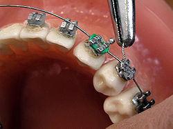
Connecting the arch-wire on brackets with wire
Occupation
Names Orthodontist
Occupation type
Specialty
Activity sectors
Dentistry
Description
Education required
Dental degree, specialty training
Fields of
employment
Private practices, hospitals

Orthodontics[a][b] is a dentistry specialty that addresses the diagnosis, prevention, management, and correction of mal-positioned teeth and jaws, as well as misaligned bite patterns.[2] It may also address the modification of facial growth, known as dentofacial orthopedics.

Abnormal alignment of the teeth and jaws is very common. The approximate worldwide prevalence of malocclusion was as high as 56%.[3] However, conclusive scientific evidence for the health benefits of orthodontic treatment is lacking, although patients with completed treatment have reported a higher quality of life than that of untreated patients undergoing orthodontic treatment.[4][5] The main reason for the prevalence of these malocclusions is diets with less fresh fruit and vegetables and overall softer foods in childhood, causing smaller jaws with less room for the teeth to erupt.[6] Treatment may require several months to a few years and entails using dental braces and other appliances to gradually adjust tooth position and jaw alignment. In cases where the malocclusion is severe, jaw surgery may be incorporated into the treatment plan. Treatment usually begins before a person reaches adulthood, insofar as pre-adult bones may be adjusted more easily before adulthood.

History

[edit]

Though it was rare until the Industrial Revolution,[7] there is evidence of the issue of overcrowded, irregular, and protruding teeth afflicting individuals. Evidence from Greek and Etruscan materials suggests that attempts to treat this disorder date back to 1000 BC, showcasing primitive yet impressively well-crafted orthodontic appliances. In the 18th and 19th centuries, a range of devices for the "regulation" of teeth were described by various dentistry authors who occasionally put them into practice.[8] As a modern science, orthodontics dates back to the mid-1800s.[9] The field's influential contributors include Norman William Kingsley[9] (1829–1913) and Edward Angle[10] (1855–1930). Angle created the first basic system for classifying malocclusions, a system that remains in use today.[9]

Beginning in the mid-1800s, Norman Kingsley published Oral Deformities, which is now credited as one of the first works to begin systematically documenting orthodontics. Being a major presence in American dentistry during the latter half of the 19th century, not only was Kingsley one of the early users of extraoral force to correct protruding teeth, but he was also one of the pioneers for treating cleft palates and associated issues. During the era of orthodontics under Kingsley and his colleagues, the treatment was focused on straightening teeth and creating facial harmony. Ignoring occlusal relationships, it was typical to remove teeth for a variety of dental issues, such as malalignment or overcrowding. The concept of an intact dentition was not widely appreciated in those days, making bite correlations seem irrelevant.[8]

In the late 1800s, the concept of occlusion was essential for creating reliable prosthetic replacement teeth. This idea was further refined and ultimately applied in various ways when dealing with healthy dental structures as well. As these concepts of prosthetic occlusion progressed, it became an invaluable tool for dentistry.[8]

It was in 1890 that the work and impact of Dr. Edwards H. Angle began to be felt, with his contribution to modern orthodontics particularly noteworthy. Initially focused on prosthodontics, he taught in Pennsylvania and Minnesota before directing his attention towards dental occlusion and the treatments needed to maintain it as a normal condition, thus becoming known as the "father of modern orthodontics".[8]

By the beginning of the 20th century, orthodontics had become more than just the straightening of crooked teeth. The concept of ideal occlusion, as postulated by Angle and incorporated into a classification system, enabled a shift towards treating malocclusion, which is any deviation from normal occlusion.[8] Having a full set of teeth on both arches was highly sought after in orthodontic treatment due to the need for exact relationships between them. Extraction as an orthodontic procedure was heavily opposed by Angle and those who followed him. As occlusion became the key priority, facial proportions and aesthetics were neglected. To achieve ideal occlusals without using external forces, Angle postulated that having perfect occlusion was the best way to gain optimum facial aesthetics.[8]

With the passing of time, it became quite evident that even an exceptional occlusion was not suitable when considered from an aesthetic point of view. Not only were there issues related to aesthetics, but it usually proved impossible to keep a precise occlusal relationship achieved by forcing teeth together over extended durations with the use of robust elastics, something Angle and his students had previously suggested. Charles Tweed[11] in America and Raymond Begg[12] in Australia (who both studied under Angle) re-introduced dentistry extraction into orthodontics during the 1940s and 1950s so they could improve facial esthetics while also ensuring better stability concerning occlusal relationships.[13]

In the postwar period, cephalometric radiography[14] started to be used by orthodontists for measuring changes in tooth and jaw position caused by growth and treatment.[15] The x-rays showed that many Class II and III malocclusions were due to improper jaw relations as opposed to misaligned teeth. It became evident that orthodontic therapy could adjust mandibular development, leading to the formation of functional jaw orthopedics in Europe and extraoral force measures in the US. These days, both functional appliances and extraoral devices are applied around the globe with the aim of amending growth patterns and forms. Consequently, pursuing true, or at least improved, jaw relationships had become the main objective of treatment by the mid-20th century.[8]

At the beginning of the twentieth century, orthodontics was in need of an upgrade. The American Journal of Orthodontics was created for this purpose in 1915; before it, there were no scientific objectives to follow, nor any precise classification system and brackets that lacked features.[16]

Until the mid-1970s, braces were made by wrapping metal around each tooth.[9] With advancements in adhesives, it became possible to instead bond metal brackets to the teeth.[9]

In 1972, Lawrence F. Andrews gave an insightful definition of the ideal occlusion in permanent teeth. This has had meaningful effects on orthodontic treatments that are administered regularly,[16] and these are: 1. Correct interarchal relationships 2. Correct crown angulation (tip) 3. Correct crown inclination (torque) 4. No rotations 5. Tight contact points 6. Flat Curve of Spee (0.0–2.5 mm),[17] and based on these principles, he discovered a treatment system called the straight-wire appliance system, or the pre-adjusted edgewise system. Introduced in 1976, Larry Andrews' pre-adjusted edgewise appliance, more commonly known as the straight wire appliance, has since revolutionized fixed orthodontic treatment. The advantage of the design lies in its bracket and archwire combination, which requires only minimal wire bending from the orthodontist or clinician. It's aptly named after this feature: the angle of the slot and thickness of the bracket base ultimately determine where each tooth is situated with little need for extra manipulation.[18][19][20]

Prior to the invention of a straight wire appliance, orthodontists were utilizing a non-programmed standard edgewise fixed appliance system, or Begg's pin and tube system. Both of these systems employed identical brackets for each tooth and necessitated the bending of an archwire in three planes for locating teeth in their desired positions, with these bends dictating ultimate placements.[18]

Evolution of the current orthodontic appliances

[edit]

When it comes to orthodontic appliances, they are divided into two types: removable and fixed. Removable appliances can be taken on and off by the patient as required. On the other hand, fixed appliances cannot be taken off as they remain bonded to the teeth during treatment.

Fixed appliances

[edit]

Fixed orthodontic appliances are predominantly derived from the edgewise appliance approach, which typically begins with round wires before transitioning to rectangular archwires for improving tooth alignment. These rectangluar wires promote precision in the positioning of teeth following initial treatment. In contrast to the Begg appliance, which was based solely on round wires and auxiliary springs, the Tip-Edge system emerged in the early 21st century. This innovative technology allowed for the utilization of rectangular archwires to precisely control tooth movement during the finishing stages after initial treatment with round wires. Thus, almost all modern fixed appliances can be considered variations on this edgewise appliance system.

Early 20th-century orthodontist Edward Angle made a major contribution to the world of dentistry. He created four distinct appliance systems that have been used as the basis for many orthodontic treatments today, barring a few exceptions. They are E-arch, pin and tube, ribbon arch, and edgewise systems.

E-arch

[edit]

Edward H. Angle made a significant contribution to the dental field when he released the 7th edition of his book in 1907, which outlined his theories and detailed his technique. This approach was founded upon the iconic "E-Arch" or 'the-arch' shape as well as inter-maxillary elastics.[21] This device was different from any other appliance of its period as it featured a rigid framework to which teeth could be tied effectively in order to recreate an arch form that followed pre-defined dimensions.[22] Molars were fitted with braces, and a powerful labial archwire was positioned around the arch. The wire ended in a thread, and to move it forward, an adjustable nut was used, which allowed for an increase in circumference. By ligation, each individual tooth was attached to this expansive archwire.[8]

Pin and tube appliance

[edit]

Due to its limited range of motion, Angle was unable to achieve precise tooth positioning with an E-arch. In order to bypass this issue, he started using bands on other teeth combined with a vertical tube for each individual tooth. These tubes held a soldered pin, which could be repositioned at each appointment in order to move them in place.[8] Dubbed the "bone-growing appliance", this contraption was theorized to encourage healthier bone growth due to its potential for transferring force directly to the roots.[23] However, implementing it proved troublesome in reality.

Ribbon arch

[edit]

Realizing that the pin and tube appliance was not easy to control, Angle developed a better option, the ribbon arch, which was much simpler to use. Most of its components were already prepared by the manufacturer, so it was significantly easier to manage than before. In order to attach the ribbon arch, the occlusal area of the bracket was opened. Brackets were only added to eight incisors and mandibular canines, as it would be impossible to insert the arch into both horizontal molar tubes and the vertical brackets of adjacent premolars. This lack of understanding posed a considerable challenge to dental professionals; they were unable to make corrections to an excessive Spee curve in bicuspid teeth.[24] Despite the complexity of the situation, it was necessary for practitioners to find a resolution. Unparalleled to its counterparts, what made the ribbon arch instantly popular was that its archwire had remarkable spring qualities and could be utilized to accurately align teeth that were misaligned. However, a major drawback of this device was its inability to effectively control root position since it did not have enough resilience to generate the torque movements required for setting roots in their new place.[8]

Edgewise appliance

[edit]

In an effort to rectify the issues with the ribbon arch, Angle shifted the orientation of its slot from vertical, instead making it horizontal. In addition, he swapped out the wire and replaced it with a precious metal wire that was rotated by 90 degrees in relation—henceforth known as Edgewise.[25] Following extensive trials, it was concluded that dimensions of 22 × 28 mils were optimal for obtaining excellent control over crown and root positioning across all three planes of space.[26] After debuting in 1928, this appliance quickly became one of the mainstays for multibanded fixed therapy, although ribbon arches continued to be utilized for another decade or so beyond this point too.[8]

Labiolingual

[edit]

Prior to Angle, the idea of fitting attachments on individual teeth had not been thought of, and in his lifetime, his concern for precisely positioning each tooth was not highly appraised. In addition to using fingersprings for repositioning teeth with a range of removable devices, two main appliance systems were very popular in the early part of the 20th century. Labiolingual appliances use bands on the first molars joined with heavy lingual and labial archwires affixed with soldered fingersprings to shift single teeth.

Twin wire

[edit]

Utilizing bands around both incisors and molars, a twin-wire appliance was designed to provide alignment between these teeth. Constructed with two 10-mil steel archwires, its delicate features were safeguarded by lengthy tubes stretching from molars towards canines. Despite its efforts, it had limited capacity for movement without further modifications, rendering it obsolete in modern orthodontic practice.

Begg's Appliance

[edit]

Returning to Australia in the 1920s, the renowned orthodontist, Raymond Begg, applied his knowledge of ribbon arch appliances, which he had learned from the Angle School. On top of this, Begg recognized that extracting teeth was sometimes vital for successful outcomes and sought to modify the ribbon arch appliance to provide more control when dealing with root positioning. In the late 1930s, Begg developed his adaptation of the appliance, which took three forms. Firstly, a high-strength 16-mil round stainless steel wire replaced the original precious metal ribbon arch. Secondly, he kept the same ribbon arch bracket but inverted it so that it pointed toward the gums instead of away from them. Lastly, auxiliary springs were added to control root movement. This resulted in what would come to be known as the Begg Appliance. With this design, friction was decreased since contact between wire and bracket was minimal, and binding was minimized due to tipping and uprighting being used for anchorage control, which lessened contact angles between wires and corners of the bracket.

Tip-Edge System

[edit]

Begg's influence is still seen in modern appliances, such as Tip-Edge brackets. This type of bracket incorporates a rectangular slot cutaway on one side to allow for crown tipping with no incisal deflection of an archwire, allowing teeth to be tipped during space closure and then uprighted through auxiliary springs or even a rectangular wire for torque purposes in finishing. At the initial stages of treatment, small-diameter steel archwires should be used when working with Tip-Edge brackets.

Contemporary edgewise systems

[edit]

Throughout time, there has been a shift in which appliances are favored by dentists. In particular, during the 1960s, when it was introduced, the Begg appliance gained wide popularity due to its efficiency compared to edgewise appliances of that era; it could produce the same results with less investment on the dentist's part. Nevertheless, since then, there have been advances in technology and sophistication in edgewise appliances, which led to the opposite conclusion: nowadays, edgewise appliances are more efficient than the Begg appliance, thus explaining why it is commonly used.

Automatic rotational control

[edit]

At the beginning, Angle attached eyelets to the edges of archwires so that they could be held with ligatures and help manage rotations. Now, however, no extra ligature is needed due to either twin brackets or single brackets that have added wings touching underneath the wire (Lewis or Lang brackets). Both types of brackets simplify the process of obtaining moments that control movements along a particular plane of space.

Alteration in bracket slot dimensions

[edit]

In modern dentistry, two types of edgewise appliances exist: the 18- and 22-slot varieties. While these appliances are used differently, the introduction of a 20-slot device with more precise features has been considered but not pursued yet.[27]

Straight-wire bracket prescriptions

[edit]

Rather than rely on the same bracket for all teeth, L.F. Andrews found a way to make different brackets for each tooth in the 1980s, thanks to the increased convenience of bonding.[28] This adjustment enabled him to avoid having multiple bends in archwires that would have been needed to make up for variations in tooth anatomy. Ultimately, this led to what was termed a "straight-wire appliance" system – an edgewise appliance that greatly enhanced its efficiency.[29] The modern edgewise appliance has slightly different construction than the original one. Instead of relying on faciolingual bends to accommodate variations among teeth, each bracket has a correspondingly varying base thickness depending on the tooth it is intended for. However, due to individual differences between teeth, this does not completely eliminate the need for compensating bends.[30] Accurately placing the roots of many teeth requires angling brackets in relation to the long axis of the tooth. Traditionally, this mesiodistal root positioning necessitated using second-order, or tip, bends along the archwire. However, angling the bracket or bracket slot eliminates this need for bends.

Given the discrepancies in inclination of facial surfaces across individual teeth, placing a twist, otherwise known as third-order or torque bends, into segments of each rectangular archwire was initially required with the edgewise appliance. These bends were necessary for all patients and wires, not just to avoid any unintentional movement of suitably placed teeth or when moving roots facially or lingually. Angulation of either brackets or slots can minimize the need for second-order or tip bends on archwires. Contemporary edgewise appliances come with brackets designed to adjust for any facial inclinations, thereby eliminating or reducing any third-order bends. These brackets already have angulation and torque values built in so that each rectangluar archwire can be contorted to form a custom fit without inadvertently shifting any correctly positioned teeth. Without bracket angulation and torque, second-order or tip bends would still be required on each patient's archwire.

Methods

[edit]
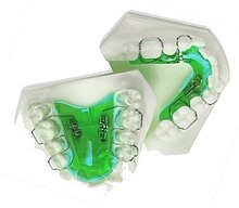
Upper and lower jaw functional expanders

A typical treatment for incorrectly positioned teeth (malocclusion) takes from one to two years, with braces being adjusted every four to 10 weeks by orthodontists,[31] while university-trained dental specialists are versed in the prevention, diagnosis, and treatment of dental and facial irregularities. Orthodontists offer a wide range of treatment options to straighten crooked teeth, fix irregular bites, and align the jaws correctly.[32] There are many ways to adjust malocclusion. In growing patients, there are more options to treat skeletal discrepancies, either by promoting or restricting growth using functional appliances, orthodontic headgear, or a reverse pull facemask. Most orthodontic work begins in the early permanent dentition stage before skeletal growth is completed. If skeletal growth has completed, jaw surgery is an option. Sometimes teeth are extracted to aid the orthodontic treatment (teeth are extracted in about half of all the cases, most commonly the premolars).[33]

Orthodontic therapy may include the use of fixed or removable appliances. Most orthodontic therapy is delivered using appliances that are fixed in place,[34] for example, braces that are adhesively bonded to the teeth. Fixed appliances may provide greater mechanical control of the teeth; optimal treatment outcomes are improved by using fixed appliances.

Fixed appliances may be used, for example, to rotate teeth if they do not fit the arch shape of the other teeth in the mouth, to adjust multiple teeth to different places, to change the tooth angle of teeth, or to change the position of a tooth's root. This treatment course is not preferred where a patient has poor oral hygiene, as decalcification, tooth decay, or other complications may result. If a patient is unmotivated (insofar as treatment takes several months and requires commitment to oral hygiene), or if malocclusions are mild.

The biology of tooth movement and how advances in gene therapy and molecular biology technology may shape the future of orthodontic treatment.[35]

Braces

[edit]
Dental braces

Braces are usually placed on the front side of the teeth, but they may also be placed on the side facing the tongue (called lingual braces). Brackets made out of stainless steel or porcelain are bonded to the center of the teeth using an adhesive. Wires are placed in a slot in the brackets, which allows for controlled movement in all three dimensions.

Apart from wires, forces can be applied using elastic bands,[36] and springs may be used to push teeth apart or to close a gap. Several teeth may be tied together with ligatures, and different kinds of hooks can be placed to allow for connecting an elastic band.[37][36]

Clear aligners are an alternative to braces, but insufficient evidence exists to determine their effectiveness.[38]

Treatment duration

[edit]

The time required for braces varies from person to person as it depends on the severity of the problem, the amount of room available, the distance the teeth must travel, the health of the teeth, gums, and supporting bone, and how closely the patient follows instructions. On average, however, once the braces are put on, they usually remain in place for one to three years. After braces are removed, most patients will need to wear a retainer all the time for the first six months, then only during sleep for many years.[39]

Headgear

[edit]

Orthodontic headgear, sometimes referred to as an "extra-oral appliance", is a treatment approach that requires the patient to have a device strapped onto their head to help correct malocclusion—typically used when the teeth do not align properly. Headgear is most often used along with braces or other orthodontic appliances. While braces correct the position of teeth, orthodontic headgear—which, as the name suggests, is worn on or strapped onto the patient's head—is most often added to orthodontic treatment to help alter the alignment of the jaw, although there are some situations in which such an appliance can help move teeth, particularly molars.

Full orthodontic headgear with headcap, fitting straps, facebow, and elastics

Whatever the purpose, orthodontic headgear works by exerting tension on the braces via hooks, a facebow, coils, elastic bands, metal orthodontic bands, and other attachable appliances directly into the patient's mouth. It is most effective for children and teenagers because their jaws are still developing and can be easily manipulated. (If an adult is fitted with headgear, it is usually to help correct the position of teeth that have shifted after other teeth have been extracted.) Thus, headgear is typically used to treat a number of jaw alignment or bite problems, such as overbite and underbite.[40]

Palatal expansion

[edit]

Palatal expansion can be best achieved using a fixed tissue-borne appliance. Removable appliances can push teeth outward but are less effective at maxillary sutural expansion. The effects of a removable expander may look the same as they push teeth outward, but they should not be confused with actually expanding the palate. Proper palate expansion can create more space for teeth as well as improve both oral and nasal airflow.[41]

Jaw surgery

[edit]

Jaw surgery may be required to fix severe malocclusions.[42] The bone is broken during surgery and stabilized with titanium (or bioresorbable) plates and screws to allow for healing to take place.[43] After surgery, regular orthodontic treatment is used to move the teeth into their final position.[44]

During treatment

[edit]

To reduce pain during the orthodontic treatment, low-level laser therapy (LLLT), vibratory devices, chewing adjuncts, brainwave music, or cognitive behavioral therapy can be used. However, the supporting evidence is of low quality, and the results are inconclusive.[45]

Post treatment

[edit]

After orthodontic treatment has been completed, there is a tendency for teeth to return, or relapse, back to their pre-treatment positions. Over 50% of patients have some reversion to pre-treatment positions within 10 years following treatment.[46] To prevent relapse, the majority of patients will be offered a retainer once treatment has been completed and will benefit from wearing their retainers. Retainers can be either fixed or removable.

Removable retainers

[edit]

Removable retainers are made from clear plastic, and they are custom-fitted for the patient's mouth. It has a tight fit and holds all of the teeth in position. There are many types of brands for clear retainers, including Zendura Retainer, Essix Retainer, and Vivera Retainer.[47] A Hawley retainer is also a removable orthodontic appliance made from a combination of plastic and metal that is custom-molded to fit the patient's mouth. Removable retainers will be worn for different periods of time, depending on the patient's need to stabilize the dentition.[48]

Fixed retainers

[edit]

Fixed retainers are a simple wire fixed to the tongue-facing part of the incisors using dental adhesive and can be specifically useful to prevent rotation in incisors. Other types of fixed retainers can include labial or lingual braces, with brackets fixed to the teeth.[48]

Clear aligners

[edit]

Clear aligners are another form of orthodontics commonly used today, involving removable plastic trays. There has been controversy about the effectiveness of aligners such as Invisalign or Byte; some consider them to be faster and more freeing than the alternatives.[49]

Training

[edit]

There are several specialty areas in dentistry, but the specialty of orthodontics was the first to be recognized within dentistry.[50] Specifically, the American Dental Association recognized orthodontics as a specialty in the 1950s.[50] Each country has its own system for training and registering orthodontic specialists.

Australia

[edit]

In Australia, to obtain an accredited three-year full-time university degree in orthodontics, one will need to be a qualified dentist (complete an AHPRA-registered general dental degree) with a minimum of two years of clinical experience. There are several universities in Australia that offer orthodontic programs: the University of Adelaide, the University of Melbourne, the University of Sydney, the University of Queensland, the University of Western Australia, and the University of Otago.[51] Orthodontic courses are accredited by the Australian Dental Council and reviewed by the Australian Society of Orthodontists (ASO). Prospective applicants should obtain information from the relevant institution before applying for admission.[52] After completing a degree in orthodontics, specialists are required to be registered with the Australian Health Practitioner Regulation Agency (AHPRA) in order to practice.[53][54]

Bangladesh

[edit]

Dhaka Dental College in Bangladesh is one of the many schools recognized by the Bangladesh Medical and Dental Council (BM&DC) that offer post-graduation orthodontic courses.[55][56] Before applying to any post-graduation training courses, an applicant must have completed the Bachelor of Dental Surgery (BDS) examination from any dental college.[55] After application, the applicant must take an admissions test held by the specific college.[55] If successful, selected candidates undergo training for six months.[57]

Canada

[edit]

In Canada, obtaining a dental degree, such as a Doctor of Dental Surgery (DDS) or Doctor of Medical Dentistry (DMD), would be required before being accepted by a school for orthodontic training.[58] Currently, there are 10 schools in the country offering the orthodontic specialty.[58] Candidates should contact the individual school directly to obtain the most recent pre-requisites before entry.[58] The Canadian Dental Association expects orthodontists to complete at least two years of post-doctoral, specialty training in orthodontics in an accredited program after graduating from their dental degree.

United States

[edit]

Similar to Canada, there are several colleges and universities in the United States that offer orthodontic programs. Every school has a different enrollment process, but every applicant is required to have graduated with a DDS or DMD from an accredited dental school.[59][60] Entrance into an accredited orthodontics program is extremely competitive and begins by passing a national or state licensing exam.[61]

The program generally lasts for two to three years, and by the final year, graduates are required to complete the written American Board of Orthodontics (ABO) exam.[61] This exam is also broken down into two components: a written exam and a clinical exam.[61] The written exam is a comprehensive exam that tests for the applicant's knowledge of basic sciences and clinical concepts.[61] The clinical exam, however, consists of a Board Case Oral Examination (BCOE), a Case Report Examination (CRE), and a Case Report Oral Examination (CROE).[61] Once certified, certification must then be renewed every ten years.[61] Orthodontic programs can award a Master of Science degree, a Doctor of Science degree, or a Doctor of Philosophy degree, depending on the school and individual research requirements.[62]

United Kingdom

[edit]

Throughout the United Kingdom, there are several Orthodontic Specialty Training Registrar posts available.[63] The program is full-time for three years, and upon completion, trainees graduate with a degree at the Masters or Doctorate level.[63] Training may take place within hospital departments that are linked to recognized dental schools.[63] Obtaining a Certificate of Completion of Specialty Training (CCST) allows an orthodontic specialist to be registered under the General Dental Council (GDC).[63] An orthodontic specialist can provide care within a primary care setting, but to work at a hospital as an orthodontic consultant, higher-level training is further required as a post-CCST trainee.[63] To work within a university setting as an academic consultant, completing research toward obtaining a Ph.D. is also required.[63]

See also

[edit]
  • Orthodontic technology
  • Orthodontic indices
  • List of orthodontic functional appliances
  • Molar distalization
  • Mouth breathing
  • Obligate nasal breathing

Notes

[edit]
  1. ^ Also referred to as orthodontia
  2. ^ "Orthodontics" comes from the Greek orthos ('correct, straight') and -odont- ('tooth').[1]

References

[edit]
  1. ^ "Definition of orthodontics | Dictionary.com". www.dictionary.com. Retrieved 2019-08-28.
  2. ^ "What is orthodontics?// Useful Resources: FAQ and Downloadable eBooks". Orthodontics Australia. Retrieved 2020-08-13.
  3. ^ Lombardo G, Vena F, Negri P, Pagano S, Barilotti C, Paglia L, Colombo S, Orso M, Cianetti S (June 2020). "Worldwide prevalence of malocclusion in the different stages of dentition: A systematic review and meta-analysis". Eur J Paediatr Dent. 21 (2): 115–22. doi:10.23804/ejpd.2020.21.02.05. PMID 32567942.
  4. ^ Whitcomb I (2020-07-20). "Evidence and Orthodontics: Does Your Child Really Need Braces?". Undark Magazine. Retrieved 2020-07-27.
  5. ^ "Controversial report finds no proof that dental braces work". British Dental Journal. 226 (2): 91. 2019-01-01. doi:10.1038/sj.bdj.2019.65. ISSN 1476-5373. S2CID 59222957.
  6. ^ von Cramon-Taubadel N (December 2011). "Global human mandibular variation reflects differences in agricultural and hunter-gatherer subsistence strategies". Proceedings of the National Academy of Sciences of the United States of America. 108 (49): 19546–19551. Bibcode:2011PNAS..10819546V. doi:10.1073/pnas.1113050108. PMC 3241821. PMID 22106280.
  7. ^ Rose, Jerome C.; Roblee, Richard D. (June 2009). "Origins of dental crowding and malocclusions: an anthropological perspective". Compendium of Continuing Education in Dentistry (Jamesburg, N.J.: 1995). 30 (5): 292–300. ISSN 1548-8578. PMID 19514263.
  8. ^ a b c d e f g h i j k Proffit WR, Fields Jr HW, Larson BE, Sarver DM (2019). Contemporary orthodontics (Sixth ed.). Philadelphia, PA. ISBN 978-0-323-54387-3. OCLC 1089435881.cite book: CS1 maint: location missing publisher (link)
  9. ^ a b c d e "A Brief History of Orthodontic Braces – ArchWired". www.archwired.com. 17 July 2019.[self-published source]
  10. ^ Peck S (November 2009). "A biographical portrait of Edward Hartley Angle, the first specialist in orthodontics, part 1". The Angle Orthodontist. 79 (6): 1021–1027. doi:10.2319/021009-93.1. PMID 19852589.
  11. ^ "The Application of the Principles of the Edge- wise Arch in the Treatment of Malocclusions: II.*". meridian.allenpress.com. Retrieved 2023-02-07.
  12. ^ "British Orthodontic Society > Museum and Archive > Collection > Fixed Appliances > Begg". www.bos.org.uk. Retrieved 2023-02-07.
  13. ^ Safirstein D (August 2015). "P. Raymond Begg". American Journal of Orthodontics and Dentofacial Orthopedics. 148 (2): 206. doi:10.1016/j.ajodo.2015.06.005. PMID 26232825.
  14. ^ Higley LB (August 1940). "Lateral head roentgenograms and their relation to the orthodontic problem". American Journal of Orthodontics and Oral Surgery. 26 (8): 768–778. doi:10.1016/S0096-6347(40)90331-3. ISSN 0096-6347.
  15. ^ Themes UF (2015-01-12). "14: Cephalometric radiography". Pocket Dentistry. Retrieved 2023-02-07.
  16. ^ a b Andrews LF (December 2015). "The 6-elements orthodontic philosophy: Treatment goals, classification, and rules for treating". American Journal of Orthodontics and Dentofacial Orthopedics. 148 (6): 883–887. doi:10.1016/j.ajodo.2015.09.011. PMID 26672688.
  17. ^ Andrews LF (September 1972). "The six keys to normal occlusion". American Journal of Orthodontics. 62 (3): 296–309. doi:10.1016/s0002-9416(72)90268-0. PMID 4505873. S2CID 8039883.
  18. ^ a b Themes UF (2015-01-01). "31 The straight wire appliance". Pocket Dentistry. Retrieved 2023-02-07.
  19. ^ Andrews LF (July 1979). "The straight-wire appliance". British Journal of Orthodontics. 6 (3): 125–143. doi:10.1179/bjo.6.3.125. PMID 297458. S2CID 33259729.
  20. ^ Phulari B (2013), "Andrews' Straight Wire Appliance", History of Orthodontics, Jaypee Brothers Medical Publishers (P) Ltd., p. 98, doi:10.5005/jp/books/12065_11, ISBN 9789350904718, retrieved 2023-02-07
  21. ^ Angle EH. Treatment of malocclusion of the teeth. 7th éd. Philadelphia: S.S.White Dental Mfg Cy, 1907
  22. ^ Philippe J (March 2008). "How, why, and when was the edgewise appliance born?". Journal of Dentofacial Anomalies and Orthodontics. 11 (1): 68–74. doi:10.1051/odfen/20084210113. ISSN 2110-5715.
  23. ^ Angle EH (1912). "Evolution of orthodontia. Recent developments". Dental Cosmos. 54: 853–867.
  24. ^ Brodie AG (1931). "A discussion on the Newest Angle Mechanism". The Angle Orthodontist. 1: 32–38.
  25. ^ Angle EH (1928). "The latest and best in Orthodontic Mechanism". Dental Cosmos. 70: 1143–1156.
  26. ^ Brodie AG (1956). "Orthodontic Concepts Prior to the Death of Edward Angle". The Angle Orthodontist. 26: 144–155.
  27. ^ Matasa CG, Graber TM (April 2000). "Angle, the innovator, mechanical genius, and clinician". American Journal of Orthodontics and Dentofacial Orthopedics. 117 (4): 444–452. doi:10.1016/S0889-5406(00)70164-8. PMID 10756270.
  28. ^ Andrews LF. Straight Wire: The Concept and Appliance. San Diego: LA Wells; 1989.
  29. ^ Andrews LF (1989). Straight wire: the concept and appliance. Lisa Schirmer. San Diego, CA. ISBN 978-0-9616256-0-3. OCLC 22808470.cite book: CS1 maint: location missing publisher (link)
  30. ^ Roth RH (November 1976). "Five year clinical evaluation of the Andrews straight-wire appliance". Journal of Clinical Orthodontics. 10 (11): 836–50. PMID 1069735.
  31. ^ Fleming PS, Fedorowicz Z, Johal A, El-Angbawi A, Pandis N, et al. (The Cochrane Collaboration) (June 2015). "Surgical adjunctive procedures for accelerating orthodontic treatment". The Cochrane Database of Systematic Reviews. 2015 (6). John Wiley & Sons, Ltd.: CD010572. doi:10.1002/14651858.cd010572. PMC 6464946. PMID 26123284.
  32. ^ "What is an Orthodontist?". Orthodontics Australia. 5 December 2019.
  33. ^ Dardengo C, Fernandes LQ, Capelli Júnior J (February 2016). "Frequency of orthodontic extraction". Dental Press Journal of Orthodontics. 21 (1): 54–59. doi:10.1590/2177-6709.21.1.054-059.oar. PMC 4816586. PMID 27007762.
  34. ^ "Child Dental Health Survey 2013, England, Wales and Northern Ireland". digital.nhs.uk. Retrieved 2018-03-08.
  35. ^ Atsawasuwan P, Shirazi S (2019-04-10). "Advances in Orthodontic Tooth Movement: Gene Therapy and Molecular Biology Aspect". In Aslan BI, Uzuner FD (eds.). Current Approaches in Orthodontics. IntechOpen. doi:10.5772/intechopen.80287. ISBN 978-1-78985-181-6. Retrieved 2021-05-16.
  36. ^ a b "Elastics For Braces: Rubber Bands in Orthodontics". Orthodontics Australia. 2019-12-15. Retrieved 2020-12-13.
  37. ^ Mitchell L (2013). An Introduction to Orthodontics. Oxford Medical Publications. pp. 220–233.
  38. ^ Rossini G, Parrini S, Castroflorio T, Deregibus A, Debernardi CL (September 2015). "Efficacy of clear aligners in controlling orthodontic tooth movement: a systematic review". The Angle Orthodontist. 85 (5): 881–889. doi:10.2319/061614-436.1. PMC 8610387. PMID 25412265. S2CID 10787375. The quality level of the studies was not sufficient to draw any evidence-based conclusions.
  39. ^ "Dental Braces and Retainers".
  40. ^ Millett DT, Cunningham SJ, O'Brien KD, Benson PE, de Oliveira CM (February 2018). "Orthodontic treatment for deep bite and retroclined upper front teeth in children". The Cochrane Database of Systematic Reviews. 2 (2): CD005972. doi:10.1002/14651858.CD005972.pub4. PMC 6491166. PMID 29390172.
  41. ^ "Palate Expander". Cleveland Clinic. Retrieved October 29, 2024.
  42. ^ "Jaw Surgery". Modern Orthodontic Clinic in Sammamish & Bellevue. Retrieved 2024-10-03.
  43. ^ Agnihotry A, Fedorowicz Z, Nasser M, Gill KS, et al. (The Cochrane Collaboration) (October 2017). Zbigniew F (ed.). "Resorbable versus titanium plates for orthognathic surgery". The Cochrane Database of Systematic Reviews. 10 (10). John Wiley & Sons, Ltd: CD006204. doi:10.1002/14651858.cd006204. PMC 6485457. PMID 28977689.
  44. ^ "British Orthodontic Society > Public & Patients > Your Jaw Surgery". www.bos.org.uk. Retrieved 2019-08-28.
  45. ^ Fleming PS, Strydom H, Katsaros C, MacDonald L, Curatolo M, Fudalej P, Pandis N, et al. (Cochrane Oral Health Group) (December 2016). "Non-pharmacological interventions for alleviating pain during orthodontic treatment". The Cochrane Database of Systematic Reviews. 2016 (12): CD010263. doi:10.1002/14651858.CD010263.pub2. PMC 6463902. PMID 28009052.
  46. ^ Yu Y, Sun J, Lai W, Wu T, Koshy S, Shi Z (September 2013). "Interventions for managing relapse of the lower front teeth after orthodontic treatment". The Cochrane Database of Systematic Reviews. 2014 (9): CD008734. doi:10.1002/14651858.CD008734.pub2. PMC 10793711. PMID 24014170.
  47. ^ "Clear Retainers | Maintain Your Hard to Get Smile with Clear Retainers". Retrieved 2020-01-13.
  48. ^ a b Martin C, Littlewood SJ, Millett DT, Doubleday B, Bearn D, Worthington HV, Limones A (May 2023). "Retention procedures for stabilising tooth position after treatment with orthodontic braces". The Cochrane Database of Systematic Reviews. 2023 (5): CD002283. doi:10.1002/14651858.CD002283.pub5. PMC 10202160. PMID 37219527.
  49. ^ Putrino A, Barbato E, Galluccio G (March 2021). "Clear Aligners: Between Evolution and Efficiency-A Scoping Review". International Journal of Environmental Research and Public Health. 18 (6): 2870. doi:10.3390/ijerph18062870. PMC 7998651. PMID 33799682.
  50. ^ a b Christensen GJ (March 2002). "Orthodontics and the general practitioner". Journal of the American Dental Association. 133 (3): 369–371. doi:10.14219/jada.archive.2002.0178. PMID 11934193.
  51. ^ "How to become an orthodontist". Orthodontics Australia. 26 September 2017.
  52. ^ "Studying orthodontics". Australian Society of Orthodontists. 26 September 2017.
  53. ^ "Specialties and Specialty Fields". Australian Health Practitioners Regulation Agency.
  54. ^ "Medical Specialties and Specialty Fields". Medical Board of Australia.
  55. ^ a b c "Dhaka Dental College". Dhaka Dental College. Archived from the original on October 28, 2017. Retrieved October 28, 2017.
  56. ^ "List of recognized medical and dental colleges". Bangladesh Medical & Dental Council (BM&DC). Retrieved October 28, 2017.
  57. ^ "Orthodontic Facts - Canadian Association of Orthodontists". Canadian Association of Orthodontists. Retrieved 26 October 2017.
  58. ^ a b c "FAQ: I Want To Be An Orthodontist - Canadian Association of Orthodontists". Canadian Association of Orthodontists. Retrieved 26 October 2017.
  59. ^ "RCDC - Eligibility". The Royal College of Dentists of Canada. Archived from the original on 29 October 2019. Retrieved 26 October 2017.
  60. ^ "Accredited Orthodontic Programs - AAO Members". www.aaoinfo.org.
  61. ^ a b c d e f "About Board Certification". American Board of Orthodontists. Archived from the original on 16 February 2019. Retrieved 26 October 2017.
  62. ^ "Accredited Orthodontic Programs | AAO Members". American Association of Orthodontists. Retrieved 26 October 2017.
  63. ^ a b c d e f "Orthodontic Specialty Training in the UK" (PDF). British Orthodontic Society. Retrieved 28 October 2017.


Frequently Asked Questions

It is essential to maintain recommended follow-ups with the dentist to adjust and monitor the orthodontic appliance for any problems[2].**